Chapter: Microbiology and Immunology: Structure and Function Imune System
Lymphoid Tissues and Organs
Lymphoid Tissues and Organs
The specific immune response to antigen is of two types: (a) humoral or antibody-mediated
immunity, mediated by antibodies produced by plasma cells; and (b) cell-mediated immunity, mediated by
sensitized lymphocytes. The immune system is organized into several special
tissues, which are col-lectively termed lymphoid or immune tissues. The
tissues that have evolved to a high degree of specificity of function are
termed lymphoid organs.
Lymphoid organs include the gut-associated lymphoid tis-sues—tonsils,
Peyer’s patches, and appendix—as well as aggre-gates of lymphoid tissue in the
submucosal spaces of the respiratory and genitourinary tracts. The lymphoid
organs, based on their function, are classified into central (primary) and
peripheral (secondary) lymphoid organs.
Central (Primary) Lymphoid Organs
Central or primary lymphoid organs are the major sites for
lymphopoiesis. These organs have the ability to produce pro-genitor cells of
the lymphocytic lineage. These are the organs in which precursor lymphocytes
proliferate, develop, and differ-entiate from lymphoid stem cells to become
immunologically competent cells. The primary lymphoid organs include thymus and
bone marrow. In mammals, T cells mature in thymus and B cells in fetal liver and
bone marrow. After acquiring immu-nological competency, the lymphocytes migrate
to secondary lymphoid organs to induce appropriate immune response on exposure
to antigens.
◗ Thymus
Thymus is the first lymphoid organ to develop. It reaches its
maximal size at puberty and then atrophies, with a significant decrease in both
cortical and medullary cells and an increase in the total fat content of the
organ. The thymus is a flat, bilobed organ situated above the heart. Each lobe
is surrounded by a capsule and is divided into lobules, which are separated
from each other by strands of connective tissue called trabeculae. Each lobule
is organized into two compartments: cortex and medulla. The stroma of the organ
is composed of dendritic cells, epithelial cells, and macrophages (Fig. 16-1).
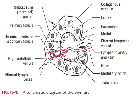
·
Cortex: It consists mainly of (a) cortical thymocytes,
theimmunologically immature T lymphocytes, and (b) a small number of macrophages and plasma cells. In addition, the
cortex contains two subpopulations of epithelial cells, the epithelial nurse
cells and the cortical epithelial cells, which form a network within the
cortex.
·
Medulla: It contains predominantly
mature T lymphocytesand has a larger epithelial cell-to-lymphocyte ratio than
the cor-tex. The concentric rings of squamous epithelial cells known as
Hassall’s corpuscles are found exclusively in the medulla.
Thymus is the site where a large diversity of T cells is produced
and so they can recognize and act against a myr-iad number of antigen–MHCs
(major histocompatibility complexes). The thymus induces the death of those T
cells that cannot recognize antigen–MHCs. It also induces death of those T
cells that react with self-antigen MHC and pose a danger of causing autoimmune
disease. More than 95% of all thymocytes die by apoptosis in the thymus without
ever reaching maturity.
◗ Bone marrow
Some lymphoid cells develop and mature within
the bone mar-row and are referred to as B cells (B for bursa of Fabricius, or
bone marrow). The function of bursa of Fabricius in birds is played by bone
marrow in humans. Bone marrow is the site for prolifera-tion of stem cells and
for the origin of pre-B cells and their matu-ration to become
immunoglobulin-producing lymphocytes.
Immature B cells proliferate and differentiate within the bone
marrow. Stromal cells within the bone marrow interact directly with the B cells
and secrete various cytokines that are required for the development of B cells.
Like thymic selection during T-cell maturation, a selection process within the
bone marrow eliminates B cells with self-reactive antibody receptors.
B lymphocytes develop their B-cell receptors (BCRs) by DNA
rearrangement. They express auxiliary molecules, such as Iga and Igb, and begin to express IgM on
their surfaces before leaving the bone marrow. Subsequently, mature B
lymphocytes also acquire C3 and Fc receptors on their surfaces. B lympho-cytes
on their surfaces either bear IgM alone or in association with IgG or IgA
depending upon the production of particular class of immunoglobulin. The B
lymphocytes are transformed into plasma cells and secrete antibodies. B
lymphocytes are primarily responsible for antibody-mediated immunity.
Peripheral (Secondary) Lymphoid Organs
Peripheral or secondary lymphoid organs consist of (a) lymph nodes, (b) spleen, and (c)
nonencapsulated structures, such as mucosa-associated lymphoid tissues (MALT).
These organs serve as the sites for interaction of mature lymphocytes with
antigens.
◗ Lymph nodes
The lymph nodes are extremely numerous and disseminated all over
the body. They play a very important and dynamic role in the initial or
inductive states of the immune response. Lymph nodes measure 1–25 mm in
diameter and are surrounded by a connective tissue capsule. The lymph node has
two main parts: cortex and medulla. The reticulum or framework of the lymph
node is composed of phagocytes and specialized types of reticular or dendritic
cells (Color Photo 10).
Cortex: The cortex and the deep cortex,
also known as para-cortical area, are densely populated by lymphocytes. Roughly
spherical areas containing densely packed lymphocytes, termed primary lymphoid
follicles or nodules, are found in the cortex. B and T lymphocytes are found in
different areas in the cortex.
The primary lymphoid follicles predominately contain B lymphocytes.
They also contain macrophages,
dendritic cells, and some T lymphocytes. The primary follicles are very densely
packed with small lymphocytes, not actively involved in an immune response. The
larger, less dense follicles, termed secondary follicles, are found in the
cortex of a lymph node draining an area in which an infection has taken place.
The secondary follicles contain clear germinal centers where B lym-phocytes actively
divide as a result of antigenic stimulation.
T
lymphocytes are found predominantly in the deep cortex or paracortical area;
for this reason, the paracortical area is des-ignated as T-dependent.
Interdigitating cells are also present in this area, where they present antigen
to T lymphocytes.
Medulla: It is less densely populated and is composed mainlyof medullary
cords. These cords are elongated branching bands of the lymphocytes, plasma
cells, and macrophages. They drain into the hilar efferent lymphatic vessels.
Plasma cells are also found in the medullary cords.
Following
the period of division, there is a rigorous selection process in which more
than 90% of these B cells die by apopto-sis or cell death. As antigen is
carried into a regional node by the lymph, it is trapped, processed, and
presented together with class II MHC molecules by interdigitating dendritic
cells in the paracortex, resulting in the activation of TH cells.
The initial activation of B cells is also thought to take place within the
T-cell-rich paracortex. Once activated, TH and B cells form small
foci consisting largely of pro-liferating B cells at the edges of the
paracortex. Some B cells within the foci differentiate into plasma cells
secreting IgM and IgG.
◗
Spleen
The spleen is the largest lymphoid organ. It is
a large, ovoid sec-ondary lymphoid organ situated high in the left abdominal
cavity. The spleen parenchyma is heterogeneous and is composed of the white and
the red pulp. It is surrounded by a capsule made up of connective tissue (Color
Photo 11). The spleen unlike the lymph nodes is not supplied by lymphatic
vessels. Instead, blood-borne antigens and lymphocytes are carried into the
spleen through the splenic artery. The narrow central arterioles, which are
derived from the splenic artery after multiple branchings, are surrounded by
lymphoid tissue (periarteriolar lymphatic sheath). In the white pulp, T
lymphocytes are found in the lymphatic sheath imme-diately surrounding the
arteriole. B lymphocytes are primarily found in perifollicular area, germinal
center, and mantle layer, which lie more peripherally relative to the
arterioles.
The effect of splenectomy on the immune response depends on the age
at which the spleen is removed:
·
In
children, splenectomy often leads to an increased incidence of bacterial sepsis
caused primarily by Streptococcus
pneumoniae, Neisseria meningitidis,
and Haemophilus influenzae.
·
In
adults, the adverse effects are less; although in some, it makes the host more
susceptible to blood-borne bacterial infections.
◗ Mucosa-associated lymphoid tissues
Mucosa-associated lymphoid tissues (MALT) consist of the lymphoid
tissues of the intestinal tract, genitourinary tract, tracheobronchial tree,
and mammary glands. All of the MALT are noncapsulated and contain both T and B
lymphocytes, and the latter predominate. Structurally, these tissues include
clus-ters of lymphoid cells in the lamina propria of intestinal villi, tonsils,
appendix, and Peyer’s patches.
Tonsils: These are present in the
oropharynx and arepredominantly populated by B lymphocytes. These are the sites
of intense antigenic stimulation, as shown by the presence of numerous
secondary follicles with germinal centers in the ton-sillar crypts.
Peyer’s patches: These are lymphoid structures
that arefound within the submucosal layer of the intestinal lining.
The follicles of the Peyer’s patches are extremely rich in B cells,
which differentiate into IgA-producing plasma cells. Specialized epithelial
cells, known as M cells, are found in abundance in the dome epithelia of
Peyer’s patches, particu-larly at the ileum. These cells take up small
particles, virus, bacteria, etc., and deliver them to submucosal macrophages,
where the engulfed material is processed and presented to T and B lymphocytes.
Related Topics