Chapter: Ophthalmology: Lacrimal System
Lacrimal System: Basic Knowledge
Lacrimal System
Basic Knowledge
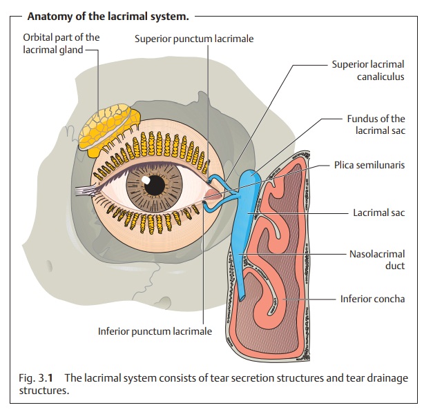
The lacrimal system (Fig. 3.1) consists of two
sections:
❖ Structures that secrete tear fluid.
❖ Structures that facilitate tear drainage.
Position, structure, and nerve supply of the lacrimal gland:
Thelacrimalgland
is about the size of a walnut;
it lies beneath the superior temporal mar-gin of the orbital bone in the
lacrimal fossa of the frontal bone and is neithervisible
nor palpable. A palpable lacrimal gland is usually a sign of a
pathologicchange such as dacryoadenitis. The tendon of the levator palpebrae
muscle divides the lacrimal gland into a larger
orbital part (two-thirds) and a smallerpalpebral
part (one-third). Several tinyaccessory
lacrimal glands (glands ofKrause and Wolfring) located in the superior
fornix secrete additional seroustear fluid.
The lacrimal gland receives its sensory supply from the lacrimal nerve.
Its parasympathetic secretomotor nerve supply comes from the nervus interme-dius. The sympathetic
fibers arise from the superior cervical sympatheticganglion and follow the
course of the blood vessels to the gland.
Tear film:
The tear film (Fig. 3.2)
that moistens the conjunctiva and cornea iscomposed of three layers:
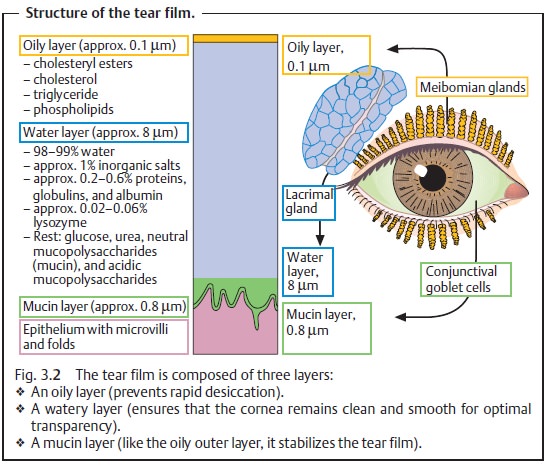
1. The outer oily layer (approximately 0.1 µm thick) is a product of the mei-bomian glands and the sebaceous glands and sweat glands of the margin of the eyelid. The primary function of this layer is to stabilize the tear film.With its hydrophobic properties, it prevents rapid evaporation like a layer of wax.
2. The middle watery layer (approximately 8 µm thick) is
produced by the lacrimal gland and
the accessory lacrimal glands (glands
of Krause andWolfring). Its task is to clean the surface of the cornea and
ensure mobility of the palpebral conjunctiva over the cornea and a smooth
corneal surface for high-quality optical
images.
3. The inner mucin layer (approximately 0.8 µm thick) is
secreted by the goblet cells of the
conjunctiva and the lacrimal gland.
It is hydrophilic withrespect to the microvilli of the corneal epithelium,
which also helps to sta-bilize the tear
film. This layer prevents the watery layer from forming beadson the cornea
and ensures that the watery layer
moistens the entire surfaceof the cornea and conjunctiva.
Lysozyme, beta-lysin, lactoferrin, and gamma
globulin (IgA) are tear-specificproteins that give the tear fluidantimicrobial characteristics.
Tear drainage:
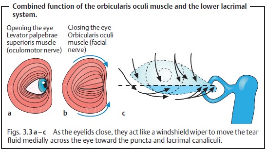
The shingle-like arrangement of thefibers of the orbicularisoculi muscle (supplied by the facial
nerve) causes the eye to close progress-ively from lateral to medial instead of
the eyelids simultaneously closing along their entire length. This windshield wiper motion moves the tear fluid
medially across the eye toward the medial canthus (Figs. 3.3a – c).
The superior and inferior puncta lacrimales collect the tears, which then drain through the superior and inferior lacrimal canaliculi into the lacrimalsac. From there they pass through the nasolacrimal duct into the inferior concha (see Fig. 3.1).
Related Topics