Organs, Structure, Functions - Human Digestive System | 9th Science : Organ Systems in Animals
Chapter: 9th Science : Organ Systems in Animals
Human Digestive System
Human Digestive System
The food we eat not only
contain simple substances like vitamins and minerals butalso contain complex
substances such as carbohydrates, proteins and fats. The body cannot use these
complex substances unless they are converted into simple substances. The ve
stages of nutrition process include ingestion, digestion, absorption, assimilation
and egestion.
The process of nutrition
begins with intake of food, called ingestion. The breakdown of large
complex insoluble food molecules into small, simpler soluble and di usible
particles by the action of digestive enzymes is called digestion. Parts
of the body concerned with the digestion of food form the digestive
system.
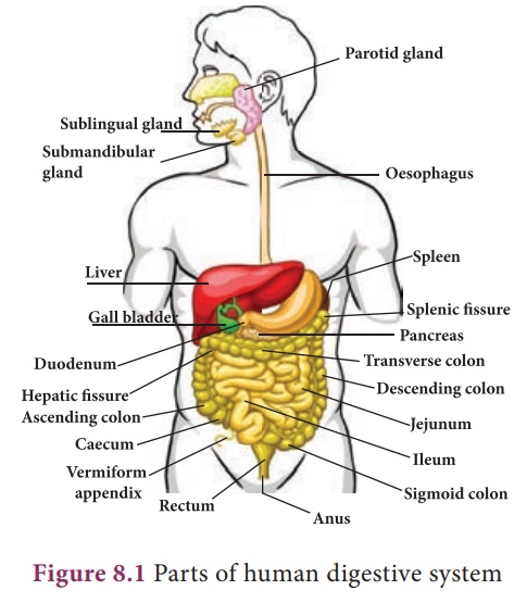
1. Organs of digestive system
The digestive system
consists of two sets of organs. They are as follows;
Alimentary canal (digestive
tract/gastro-intestinal tract) It is the passage of food starting from the
mouth and ends with the anus.
The glands associated
with the digestive system are the salivary glands, gastric glands, pancreas,
liver and intestinal glands.
2. Structure of the Alimentary Canal
Alimentary canal is a
muscular coiled, tubular structure. It consists of mouth, buccal cavity,
pharynx, oesophagus, stomach, small intestine (consisting of duodenum, jejunum
and ileum), large intestine (consisting of caecum, colon and rectum) and anus.
Mouth: The mouth leads into the
buccal cavity. It is bound by two soft, movable upper and lower lips.
The buccal cavity is a large space bound above by the palate (which
separates the wind pipe and food tube), below by the throat and on the sides by
the jaws. The jaws bear teeth.
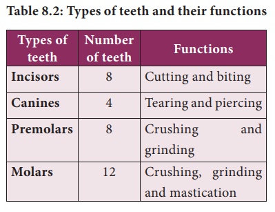
Teeth: Teeth are hard
structures meant for holding, cutting, grinding and crushing the food.
In human beings two sets of teeth (Diphyodont) are developed in their
life time. The first appearing set of 20 teeth called temporary or milk
teeth are replaced by the second set of thirty two permanent teeth, sixteen in
each jaw. Each tooth has a root fitted in the gum (Theocodont).
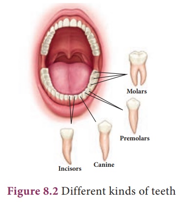
Permanent teeth are of four types (Heterodont), according to their
structure and function namely incisors, canines, premolars and
molars.
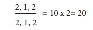
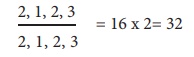
Dental formula represents the number
of different type of teeth present in each half of a jaw (upper and lower
jaw). The types of teeth are denoted as incisors (i), canine (c), premolars
(pm) and molars (m). The dental formula is presented as:
For Milk teeth in each half of upper
and lower jaw:
For Permanent teeth in each half of upper
and lower jaw:
Salivary glands: Three pairs of salivary
glands are present in the mouth cavity. They are: parotid glands,
sublingual glands and submaxillary or submandibular glands
i. Parotid glands are the largest salivary
glands, which lie in the cheeks in front of the ears (in Greek Par - near ;
otid - ear).
ii. Sublingual glands are the smallest glands and lie beneath
the tongue.
iii. Submaxillary or Submandibular
glands lie at the angles of the lower jaw.
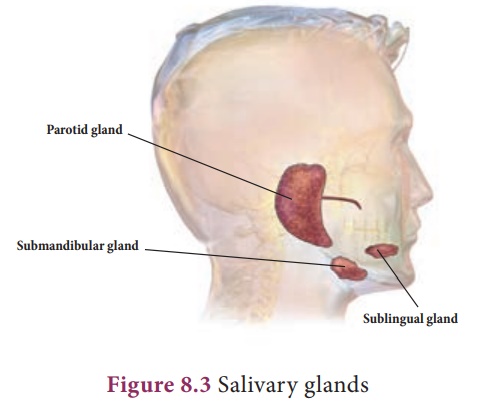
The salivary glands
secrete a viscous fluid called saliva, approximately 1.5 liters per day. It
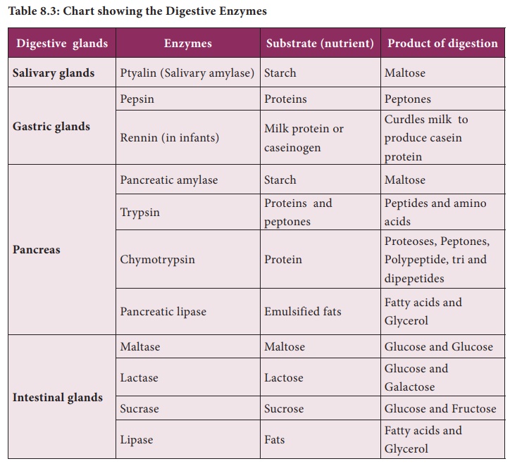
digests starch by the action of the enzyme ptyalin (amylase) in the
saliva which converts starch (polysaccharide) into maltose (disaccharide).
Saliva also contain an antibacterial enzyme called lysozyme.
Tongue: The tongue is a
muscular, sensory organ which helps in mixing the food with the saliva.
The taste buds on the tongue help to recognize the taste of food.
The masticated food in
the buccal cavity becomes a bolus which is rolled by the tongue and passed
through pharynx into the oesophagus by swallowing. During swallowing, the
epiglottis (a muscular flap-like structure at the tip of the glottis, beginning
of trachea) closes and prevents the food from entering into trachea (wind
pipe).
Pharynx
The pharynx is a
membrane lined cavity behind the nose and mouth, connecting them to the
oesophagus. It serves as a pathway for the movement of food from mouth to
oesophagus.
Oesophagus
Oesophagus or the food
pipe is a muscular-membranous canal about 22 cm in length. It conducts food
from pharynx to the stomach by peristalsis (wave-like movement) produced by the
rhythmic contraction and relaxation of the muscular walls of alimentary canal.
Stomach
The stomach is a wide
J-shaped muscular organ located between oesophagus and the small intestine. The
gastric glands present in the inner walls of the stomach secrete gastric juice.
The gastric juice is colourless, highly acidic, containing mucus, hydrochloric
acid and enzymes rennin (in infants) and pepsin.
Inactive pepsinogen is
converted to active pepsin which acts on the proteins in the ingested
food. Hydrochloric acid kills the bacteria swallowed along with food and
makes the medium acidic while the mucus protects the wall of the stomach. The
action of the gastric juice and churning of food in the stomach convert the
bolus into a semi-digested food called chyme. The chyme moves to the
intestine slowly through the pylorus.
Small intestine The small intestine is
the longest part of the alimentary canal, which is a long coiled tube
measuring about 5 – 7 m. It comprises three parts- duodenum, jejunum and ileum.
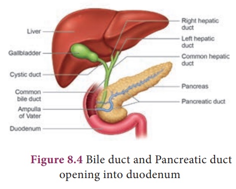
1. Duodenum is C-shaped and receives
the bile duct (from liver) and pancreatic duct (from pancreas).
2. Jejunum is the middle part of
the small intestine. It is a short region of the small intestine. The
secretion of the small intestine is intestinal juice which contains the enzymes
like sucrase, maltase, lactase and lipase.
3. Ileum forms the lower part of
the small intestine and opens into the large intestine. Ileum is the
longest part of the small intestine. It contains minute finger like projections
called villi (one millimeter in length) where absorption of food takes
place. They are approximately 4 million in number. Internally, each villus
contains fine blood capillaries and lacteal tubes,
The small intestine
serves both for digestion and absorption. It receives (i) the bile from liver
and (ii) the pancreatic juice from pancreas in the duodenum. The intestinal
glands secrete the intestinal juices.
Liver: It is the largest
digestive gland of the body which is reddish brown in colour. It is
divided into two main lobes, right and le lobes. The right lobe is larger than
the le lobe. On the under surface of the liver, gall bladder is present. The liver
cells secrete bile which is temporarily stored in the gall bladder. Bile
is released into small intestine when food enters in it. It has bile salts
(sodium glycolate and sodium tauraglycolate) and bile pigments
(bilirubin and biliviridin). Bile salts help in the digestion of fats by
bringing about their emulsi cation (conversion of large fat droplets
into small ones).
Functions of Liver
·
Controls blood sugar and amino acid levels
·
Synthesizes foetal red blood cells
·
Produces fibrinogen and prothrombin, used for clotting of blood
·
Destroys red blood cells
·
Stores iron, copper, vitamins A and D.
·
Produces heparin (an anticoagulant)
·
Excretes toxic and metallic poisons
·
Detoxifies substances including drugs and alcohol
Pancreas
It is a lobed, leaf
shaped gland situated between the stomach and duodenum. Pancreas acts
both as an exocrine gland and as an endocrine gland. The exocrine
part of the pancreatic gland secretes pancreatic juice which contains three
enzymes- lipase, trypsin and amylase which acts on fats, proteins and starch
respectively. The gland’s upper surface bears the islets of Langerhans
which have endocrine cells and secrete hormones in which α (alpha) cells
secrete glucagon and β (beta) cells secrete insulin.
The intestinal glands
secrete intestinal juice called succus entericus which contains enzymes
like maltase, lactase, sucrase and lipase which act in an alkaline medium. From
the duodenum the food is slowly moved down to ileum, where the digested food
gets absorbed
Absorption of food
Absorption is the
process by which nutrients obtained after digestion are absorbed by villi and
circulated throughout the body by blood and lymph and supplied to all body
cells according to their requirements.
Assimilation of food
Assimilation means the
incorporation of the absorbed food materials into the tissue cells as their
internal and homogenous component. The final products of fat digestion (fatty
acids and glycerol) are again converted
Chart showing the Digestive Enzymes
into fats and excess
fats are stored in adipose tissue. The excess sugars are converted into a
complex polysaccharide, glycogen in the liver. The amino acids are utilized to
synthesize different proteins required for the body.
Large intestine
The unabsorbed and
undigested food is passed into the large intestine. It extends from the ileum
to the anus. It is about 1.5 meters in length. It has three parts- caecum,
colon and rectum.
The caecum is a small
blind pouch like structure situated at the junction of the small and large
intestine. From its blind end a nger – like structure called vermiform
appendix arises. It is a vestigeal (functionless) organ in human
beings.
The colon is much
broader than ileum. It passes up the abdomen on the right (ascending colon),
crosses to the le just below the stomach (transverse colon) and
down on the le side (descending colon). The rectum

It is kept closed by a ring of muscles called anal
sphincter which opens when passing stools.
Egestion: e undigested or
unassimilated portion of the ingested food material is thrown out from
the body through the anal aperture as faecal matter. is is known as egestion
or defaecation.
Related Topics