Chapter: Medical Physiology: Cerebral Cortex, Intellectual Functions of the Brain, Learning and Memory
Functions of Specific Cortical Areas
Functions of Specific Cortical Areas
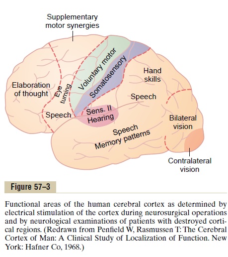
Studies in human beings by neurosurgeons, neurolo-gists, and neuropathologists have shown that different cerebral cortical areas have separate functions. Figure 57–3 is a map of some of these functions as determined by Penfield and Rasmussen from electrical stimulation of the cortex in awake patients or during neurological examination of patients after portions of the cortex had been removed. The electrically stimulated patients told their thoughts evoked by the stimulation, and sometimes they experienced movements. Occasionally they spontaneously emitted a sound or even a word or gave some other evidence of the stimulation.
Putting large amounts of information together from many different sources gives a more general map, as shown in Figure 57–4. This figure shows the major primary and secondary premotor and supplementary motor areas of the cortex as well as the major primary and secondary sensory areas for somatic sensation, vision, and hearing. The primary motor areas have direct con-nections with specific muscles for causing discrete muscle movements. The primary sensory areas detect specific sensations—visual, auditory, or somatic— transmitted directly to the brain from peripheral sensory organs.
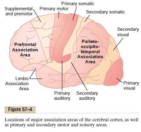
The secondary areas make sense out of the signals in the primary areas. For instance, the supplementary and premotor areas function along with the primary motor cortex and basal ganglia to provide “patterns” of motor activity. On the sensory side, the secondary sensory areas, located within a few centimeters of the primary areas, begin to analyze the meanings of the specific sensory signals, such as (1) interpretation of the shape or texture of an object in one’s hand; (2) interpretation of color, light intensity, directions of lines and angles, and other aspects of vision; and (3) interpretations of the meanings of sound tones and sequence of tones in the auditory signals.
Association Areas
Figure 57–4 also shows several large areas of the cere-bral cortex that do not fit into the rigid categories of primary and secondary motor and sensory areas.These areas are called association areas because they receive and analyze signals simultaneously from multiple regions of both the motor and sensory cortices as well as from subcortical structures. Yet even the association areas have their specializations. The most important association areas are (1) the parieto-occipitotemporalassociation area, (2) the prefrontal association area,and (3) the limbic association area. Following are explanations of the functions of these areas.
Parieto-occipitotemporal Association Area. This associationarea lies in the large parietal and occipital cortical space bounded by the somatosensory cortex anteri-orly, the visual cortex posteriorly, and the auditory cortex laterally. As would be expected, it provides a high level of interpretative meaning for signals from all the surrounding sensory areas. However, even the parieto-occipitotemporal association area has its own functional subareas, which are shown in Figure 57–5.

1. Analysis of the Spatial Coordinates of the Body. Anarea beginning in the posterior parietal cortex and extending into the superior occipital cortex provides continuous analysis of the spatial coordinates of all parts of the body as well as of the surroundings of the body. This area receives visual sensory informa-tion from the posterior occipital cortex and simulta-neous somatosensory information from the anterior parietal cortex. From all this information, it computes the coordinates of the visual, auditory, and body surroundings.
2.Area for Language Comprehension. The major areafor language comprehension, called Wernicke’s area, lies behind the primary auditory cortex in the posteriorpart of the superior gyrus of the temporal lobe. Wediscuss this area much more fully later; it is the most important region of the entire brain for higher intel-lectual function because almost all such intellectual functions are language based.
3.Area for Initial Processing of Visual Language (Reading). Posterior to the language comprehensionarea, lying mainly in the anterolateral region of the occipital lobe, is a visual association area that feeds visual information conveyed by words read from a book into Wernicke’s area, the language comprehen-sion area. This so-called angular gyrus area is needed to make meaning out of the visually perceived words. In its absence, a person can still have excellent language comprehension through hearing but not through reading.
4.Area for Naming Objects. In the most lateral por-tions of the anterior occipital lobe and posterior tem-poral lobe is an area for naming objects. The names arelearned mainly through auditory input, whereas the physical natures of the objects are learned mainly through visual input. In turn, the names are essential for both auditory and visual language comprehension (functions performed in Wernicke’s area located imme-diately superior to the auditory “names” region and anterior to the visual word processing area).
Prefrontal Association Area. We learned that the prefrontal association area functions in close association with the motor cortex to plan complex patterns and sequences of motor movements. To aid in this function, it receives strong input through a massive subcortical bundle of nerve fibers connecting the parieto-occipitotemporal association area with the prefrontal association area. Through this bundle, the prefrontal cortex receives much preanalyzed sensory information, especially information on the spatial coordinates of the body that is necessary for planning effective movements. Much of the output from the prefrontal area into the motor control system passes through the caudate portion of the basal ganglia-thalamic feedback circuit for motor planning, which provides many of the sequential and parallel compo-nents of movement stimulation.
The prefrontal association area is also essential tocarrying out “thought” processes in the mind. This pre-sumably results from some of the same capabilities of the prefrontal cortex that allow it to plan motor activ-ities. It seems to be capable of processing nonmotor as well as motor information from widespread areas of the brain and therefore to achieve nonmotor types of thinking as well as motor types. In fact, the prefrontal association area is frequently described simply as important for elaboration of thoughts, and it is said to store on a short-term basis “working memories” that are used to combine new thoughts while they are entering the brain.
Broca’s Area. A special region in the frontal cortex,called Broca’s area, provides the neural circuitry forword formation. This area, shown in Figure 57–5, islocated partly in the posterior lateral prefrontal cortex and partly in the premotor area. It is here that plans and motor patterns for expressing individual words or even short phrases are initiated and executed. This area also works in close association with Wernicke’s language comprehension center in the temporal association cortex.
An especially interesting discovery is the following: When a person has already learned one language and then learns a new language, the area in the brain where the new language is stored is slightly removed from the storage area for the first language. If both languages are learned simultaneously, they are stored together in the same area of the brain.
Limbic Association Area. Figures 57–4 and 57–5 show stillanother association area called the limbic associationarea. This area is found in the anterior pole of the tem-poral lobe, in the ventral portion of the frontal lobe, and in the cingulate gyrus lying deep in the longitu-dinal fissure on the midsurface of each cerebral hemisphere. It is concerned primarily with behavior,emotions, and motivation. We will learn that the limbic cortex is part of a much more extensive system, the limbic system, that includes a complex set of neuronal structures in the midbasal regions of the brain. This limbic system provides most of the emo-tional drives for activating other areas of the brain and even provides motivational drive for the process of learning itself.
Area for Recognition of Faces
An interesting type of brain abnormality called prosophenosia is inability to recognize faces. Thisoccurs in people who have extensive damage on the medial undersides of both occipital lobes and along the medioventral surfaces of the temporal lobes, as shown in Figure 57–6. Loss of these face recognition areas, strangely enough, results in little other abnor-mality of brain function.
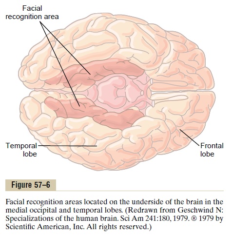
One wonders why so much of the cerebral cortex should be reserved for the simple task of face recog-nition. Most of our daily tasks involve associations with other people, and one can see the importance of this intellectual function.
The occipital portion of this facial recognition area is contiguous with the visual cortex, and the temporal portion is closely associated with the limbic system that has to do with emotions, brain activation, and control of one’s behavioral response to the environ-ment.
Comprehensive Interpretative Function of the Posterior Superior Temporal Lobe—“Wernicke’s Area” (a General Interpretative Area)
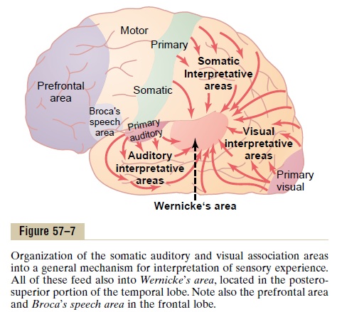
The somatic, visual, and auditory association areas all meet one another in the posterior part of the superior temporal lobe, shown in Figure 57–7, where the tem-poral, parietal, and occipital lobes all come together. This area of confluence of the different sensory inter-pretative areas is especially highly developed in the dominant side of the brain—the left side in almost allright-handed people—and it plays the greatest single role of any part of the cerebral cortex for the higher comprehension levels of brain function that we call intelligence. Therefore, this region has been called bydifferent names suggestive of an area that has almost global importance: the general interpretative area, the gnostic area, the knowing area, the tertiary association area, and so forth. It is best known as Wernicke’s area in honor of the neurologist who first described its special significance in intellectual processes.
After severe damage in Wernicke’s area, a person might hear perfectly well and even recognize different words but still be unable to arrange these words into a coherent thought. Likewise, the person may be able to read words from the printed page but be unable to recognize the thought that is conveyed.
Electrical stimulation in Wernicke’s area of a con-scious person occasionally causes a highly complex thought. This is particularly true when the stimulation electrode is passed deep enough into the brain to approach the corresponding connecting areas of the thalamus. The types of thoughts that might be experi-enced include complicated visual scenes that one might remember from childhood, auditory hallucina-tions such as a specific musical piece, or even a state-ment made by a specific person. For this reason, it is believed that activation of Wernicke’s area can call forth complicated memory patterns that involve more than one sensory modality even though most of the individual memories may be stored elsewhere. This belief is in accord with the importance of Wernicke’s area in interpreting the complicated meanings of dif-ferent patterns of sensory experiences.
Angular Gyrus—Interpretation of Visual Information. Theangular gyrus is the most inferior portion of the pos-terior parietal lobe, lying immediately behind Wer-nicke’s area and fusing posteriorly into the visual areas of the occipital lobe as well. If this region is destroyed while Wernicke’s area in the temporal lobe is still intact, the person can still interpret auditory experi-ences as usual, but the stream of visual experiences passing into Wernicke’s area from the visual cortex is mainly blocked. Therefore, the person may be able to see words and even know that they are words but not be able to interpret their meanings. This is the condi-tion called dyslexia, or word blindness.
Let us again emphasize the global importance of Wernicke’s area for processing most intellectual functions of the brain. Loss of this area in an adult usually leads thereafter to a lifetime of almost demented existence.
Concept of the Dominant Hemisphere
The general interpretative functions of Wernicke’s area and the angular gyrus, as well as the functions of the speech and motor control areas, are usually much more highly developed in one cerebral hemisphere than in the other. Therefore, this hemi-sphere is called the dominant hemisphere. In about 95 per cent of all people, the left hemisphere is the dom-inant one.
Even at birth, the area of the cortex that will even-tually become Wernicke’s area is as much as 50 per cent larger in the left hemisphere than in the right in more than one half of neonates. Therefore, it is easy to understand why the left side of the brain might become dominant over the right side. However, if for some reason this left side area is damaged or removed in very early childhood, the opposite side of the brain will usually develop dominant characteristics.
A theory that can explain the capability of one hemisphere to dominate the other hemisphere is the following.
The attention of the “mind” seems to be directed to one principal thought at a time. Presumably, because the left posterior temporal lobe at birth is usually slightly larger than the right, the left side normally begins to be used to a greater extent than the right. Thereafter, because of the tendency to direct one’s attention to the better developed region, the rate of learning in the cerebral hemisphere that gains the first start increases rapidly, whereas in the opposite, less-used side, learning remains slight. Therefore, the left side normally becomes dominant over the right.
In about 95 per cent of all people, the left temporal lobe and angular gyrus become dominant, and in the remaining 5 per cent, either both sides develop simul-taneously to have dual function, or, more rarely, the right side alone becomes highly developed, with full dominance.
As discussed later, the premotor speech area (Broca’s area), located far laterally in the intermediate frontal lobe, is also almost always domi-nant on the left side of the brain. This speech area is responsible for formation of words by exciting simul-taneously the laryngeal muscles, respiratory muscles, and muscles of the mouth.
The motor areas for controlling hands are also dominant in the left side of the brain in about 9 of 10 persons, thus causing right-handedness in most people.
Although the interpretative areas of the temporal lobe and angular gyrus, as well as many of the motor areas, are usually highly developed in only the left hemisphere, these areas receive sensory information from both hemispheres and are capable also of con-trolling motor activities in both hemispheres. For this purpose, they use mainly fiber pathways in the corpuscallosum for communication between the two hemi-spheres. This unitary, cross-feeding organization pre-vents interference between the two sides of the brain; such interference could create havoc with both mental thoughts and motor responses.
Role of Language in the Function of Wernicke’s Area and in Intellectual Functions
A major share of our sensory experience is converted into its language equivalent before being stored in the memory areas of the brain and before being processed for other intellectual purposes. For instance, when we read a book, we do not store the visual images of the printed words but instead store the words them-selves or their conveyed thoughts often in language form.
The sensory area of the dominant hemisphere for interpretation of language is Wernicke’s area, and this is closely associated with both the primary and sec-ondary hearing areas of the temporal lobe. This close relation probably results from the fact that the first introduction to language is by way of hearing. Later in life, when visual perception of language through the medium of reading develops, the visual informa-tion conveyed by written words is then presumably channeled through the angular gyrus, a visual associa-tion area, into the already developed Wernicke’s lan-guage interpretative area of the dominant temporal lobe.
Functions of the Parieto-occipitotemporal Cortex in the Nondominant Hemisphere
When Wernicke’s area in the dominant hemisphere of an adult person is destroyed, the person normally loses almost all intellectual functions associated with lan-guage or verbal symbolism, such as the ability to read, the ability to perform mathematical operations, and even the ability to think through logical problems. Many other types of interpretative capabilities, some of which use the temporal lobe and angular gyrus regions of the opposite hemisphere, are retained.
Psychological studies in patients with damage to the nondominant hemisphere have suggested that this hemisphere may be especially important for under-standing and interpreting music, nonverbal visual experiences (especially visual patterns), spatial rela-tions between the person and their surroundings, the significance of “body language” and intonations of people’s voices, and probably many somatic experi-ences related to use of the limbs and hands. Thus, even though we speak of the “dominant” hemisphere, this is primarily for language-based intellectual functions; the so-called nondominant hemisphere might actually be dominant for some other types of intelligence.
Higher Intellectual Functions of the Prefrontal Association Areas
For years, it has been taught that the prefrontal cortex is the locus of “higher intellect” in the human being, principally because the main difference between the brains of monkeys and of human beings is the great prominence of the human prefrontal areas. Yet, efforts to show that the prefrontal cortex is more important in higher intellectual functions than other portions of the brain have not been successful. Indeed, destruction of the language comprehension area in the posterior superior temporal lobe (Wernicke’s area) and the adjacent angular gyrus region in the dominant hemi-sphere causes much more harm to the intellect than does destruction of the prefrontal areas.The prefrontal areas do, however, have less definable but nevertheless important intellectual functions of their own. These functions can best be explained by describing what happens to patients in whom the prefrontal areas have become nonfunctional, as follows.
Several decades ago, before the advent of modern drugs for treating psychiatric conditions, it was found that some patients could receive significant relief from severe psychotic depression by severing the neuronal connections between the prefrontal areas of the brain and the remainder of the brain, that is, by a procedure called prefrontal lobotomy. This was done by inserting a blunt, thin-bladed knife through a small opening in the lateral frontal skull on each side of the head and slicing the brain at the back edge of the prefrontal lobes from top to bottom. Subsequent studies in these patients showed the following mental changes:
1.The patients lost their ability to solve complex problems.
2.They became unable to string together sequential tasks to reach complex goals.
3.They became unable to learn to do several parallel tasks at the same time.
4.Their level of aggressiveness was decreased, sometimes markedly, and, in general, they lost ambition.
5.Their social responses were often inappropriate for the occasion, often including loss of morals and little reticence in relation to sex and excretion.
6.The patients could still talk and comprehend language, but they were unable to carry through any long trains of thought, and their moods changed rapidly from sweetness to rage to exhilaration to madness.
7.The patients could also still perform most of the usual patterns of motor function that they had performed throughout life, but often without purpose.
From this information, let us try to piece together a coherent understanding of the function of the pre-frontal association areas.
Decreased Aggressiveness and Inappropriate Social Responses. These two characteristics probably resultfrom loss of the ventral parts of the frontal lobes on the underside of the brain. As explained earlier and shown in Figures 57–4 and 57–5, this area is part of the limbic association cortex, rather than of the prefrontal association cortex.
Inability to Progress Toward Goals or to Carry Through Sequen-tial Thoughts. We learned earlier that the prefrontal association areas have the capability of calling forth information from widespread areas of the brain and using this information to achieve deeper thought patterns for attaining goals. If these goals include motor action, so be it. If they do not, then the thought processes attain intellectual analytical goals.
Although people without prefrontal cortices can still think, they show little concerted thinking in logical sequence for longer than a few seconds or a minute or so at most. One of the results is that people without prefrontal cortices are easily distracted from theircentral theme of thought, whereas people with func-tioning prefrontal cortices can drive themselves to completion of their thought goals irrespective of distractions.
Elaboration of Thought, Prognostication, and Performance of Higher Intellectual Functions by the Prefrontal Areas—Concept of a “Working Memory.” Another function that has beenascribed to the prefrontal areas by psychologists and neurologists is elaboration of thought. This means simply an increase in depth and abstractness of the dif-ferent thoughts put together from multiple sources of information. Psychological tests have shown that pre-frontal lobectomized lower animals presented with successive bits of sensory information fail to keep track of these bits even in temporary memory, proba-bly because they are distracted so easily that they cannot hold thoughts long enough for memory storage to take place.
This ability of the prefrontal areas to keep track of many bits of information simultaneously and to cause recall of this information instantaneously as it is needed for subsequent thoughts is called the brain’s “working memory.” This could well explain the many functions of the brain that we associate with higher intelligence. In fact, studies have shown that the pre-frontal areas are divided into separate segments for storing different types of temporary memory, such as one area for storing shape and form of an object or a part of the body and another for storing movement.
By combining all these temporary bits of working memory, we have the abilities to (1) prognosticate; (2) plan for the future; (3) delay action in response to incoming sensory signals so that the sensory informa-tion can be weighed until the best course of response is decided; (4) consider the consequences of motor actions before they are performed; (5) solve compli-cated mathematical, legal, or philosophical problems; (6) correlate all avenues of information in diagnosing rare diseases; and (7) control our activities in accord with moral laws.
Function of the Brain in Communication—Language Input and Language Output
One of the most important differences between human beings and lower animals is the facility with which human beings can communicate with one another. Fur-thermore, because neurological tests can easily assess
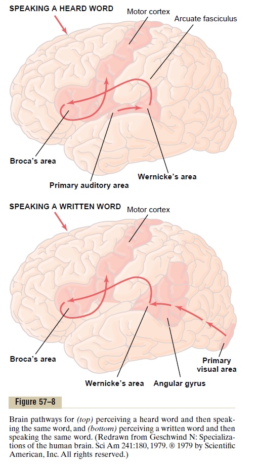
the ability of a person to communicate with others, we know more about the sensory and motor systems related to communication than about any other segment of brain cortex function. Therefore, we will review, with the help of anatomical maps of neural path-ways in Figure 57–8, function of the cortex in commu-nication. From this, one will see immediately how the principles of sensory analysis and motor control apply to this art.
There are two aspects to communication: first, the sensory aspect (language input), involving the ears andeyes, and, second, the motor aspect(language output), involving vocalization and its control.
Sensory Aspects of Communication. We noted earlier that destruction of portions of the auditory or visual association areas of the cortex can result in inabil-ity to understand the spoken word or the written word.
These effects are called, respectively, auditory receptiveaphasia and visual receptive aphasia or, more commonly, word deafness and word blindness(also called dyslexia).
Wernicke’s Aphasia and Global Aphasia. Some peopleare capable of understanding either the spoken word or the written word but are unable to interpret the thought that is expressed. This results most frequently when
Wernicke’s area in the posterior superior temporal gyrus in the dominant hemisphere is damaged or destroyed.Therefore, this type of aphasia is calledWernicke’saphasia.
When the lesion in Wernicke’s area is widespread and extends (1) backward into the angular gyrus region, (2) inferiorly into the lower areas of the temporal lobe, and (3) superiorly into the superior border of the sylvian fissure, the person is likely to be almost totally demented for language understanding or communica-tion and therefore is said to have global aphasia.
Motor Aspects of Communication. The process of speechinvolves two principal stages of mentation: (1) forma-tion in the mind of thoughts to be expressed as well as choice of words to be used and then (2) motor control of vocalization and the actual act of vocalization itself.
The formation of thoughts and even most choices of words are the function of sensory association areas of the brain. Again, it is Wernicke’s area in the posterior part of the superior temporal gyrus that is most impor-tant for this ability. Therefore, a person with either Wer-nicke’s aphasia or global aphasia is unable to formulate the thoughts that are to be communicated. Or, if the lesion is less severe, the person may be able to formu-late the thoughts but unable to put together appropri-ate sequences of words to express the thought. The person sometimes is even fluent with words but the words are jumbled.
Loss of Broca’s Area Causes Motor Aphasia. Sometimesa person is capable of deciding what he or she wants to say but cannot make the vocal system emit words instead of noises. This effect, called motor aphasia, results from damage to Broca’s speech area, which lies in the prefrontal and premotorfacial region of the cere-bral cortex—about 95 per cent of the time in the left hemisphere, as shown in Figures 57–5 and 57–8. There-fore, the skilled motor patterns for control of the larynx, lips, mouth, respiratory system, and other accessory muscles of speech are all initiated from this area.
Articulation. Finally, we have the act of articulation,which means the muscular movements of the mouth, tongue, larynx, vocal cords, and so forth that are respon-sible for the intonations, timing, and rapid changes in intensities of the sequential sounds.The facial and laryn-geal regions of the motor cortex activate these muscles,and the cerebellum, basal ganglia, and sensory cortex all help to control the sequences and intensities of muscle contractions, making liberal use of basal ganglial and cerebellar feedback mechanisms. Destruction of any of these regions can cause either total or partial inability to speak distinctly.
Summary. Figure 57–8 shows two principal pathways forcommunication. The upper half of the figure shows the pathway involved in hearing and speaking. This sequence is the following: (1) reception in the primary auditory area of the sound signals that encode the words; (2) interpretation of the words in Wernicke’s area; (3) determination, also in Wernicke’s area, of the thoughts and the words to be spoken; (4) transmission of signals from Wernicke’s area to Broca’s area by way of the arcuate fasciculus; (5) activation of the skilled motor programs in Broca’s area for control of word for-mation; and (6) transmission of appropriate signals into the motor cortex to control the speech muscles.
The lower figure illustrates the comparable steps in reading and then speaking in response.The initial recep-tive area for the words is in the primary visual area rather than in the primary auditory area. Then the infor-mation passes through early stages of interpretation in the angular gyrus region and finally reaches its full level of recognition in Wernicke’s area. From here, the sequence is the same as for speaking in response to the spoken word.
Related Topics