Chapter: Medical Physiology: Overview of the Circulation; Medical Physics of Pressure, Flow, and Resistance
Blood Pressure
Blood Pressure
Standard Units of Pressure. Blood pressure almost alwaysis measured in millimeters of mercury (mm Hg) because the mercury manometer (shown in Figure 14–7) has been used since antiquity as the standard
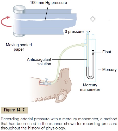
Actually, blood pres-sure means the force exerted by the blood against anyunit area of the vessel wall. When one says that thepressure in a vessel is 50 mm Hg, one means that the force exerted is sufficient to push a column of mercury against gravity up to a level 50 mm high. If the pres-sure is 100 mm Hg, it will push the column of mercury up to 100 millimeters.
Occasionally, pressure is measured in centimeters ofwater (cm H2O). A pressure of 10 cm H2O means apressure sufficient to raise a column of water against gravity to a height of 10 centimeters. One millimeter ofmercury pressure equals 1.36 cm water pressurebecause the specific gravity of mercury is 13.6 times that of water, and 1 centimeter is 10 times as great as 1 millimeter.
High-Fidelity Methods for Measuring Blood Pressure. Themercury in the mercury manometer has so much inertia that it cannot rise and fall rapidly. For this reason, the mercury manometer, although excellent for recording steady pressures, cannot respond to pressure changes that occur more rapidly than about one cycle every 2 to 3 seconds. Whenever it is desired to record rapidly changing pressures, some other type of pressure recorder is needed. Figure 14–8 demonstrates the basic principles of three electronic pressure transducers com-monly used for converting blood pressure and/or rapid changes in pressure into electrical signals and then recording the electrical signals on a high-speed electri-cal recorder. Each of these transducers uses a very thin, highly stretched metal membrane that forms one wall of the fluid chamber. The fluid chamber in turn is con-nected through a needle or catheter to the blood vessel in which the pressure is to be measured. When the pres-sure is high, the membrane bulges slightly, and when it is low, it returns toward its resting position.
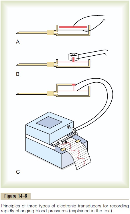
In Figure 14–8A, a simple metal plate is placed a few hundredths of a centimeter above the membrane. When the membrane bulges, the membrane comes closer to the plate, which increases the electrical capacitance between these two, and this change in capacitance can be recorded using an appropriate electronic system.
In Figure 14–8B, a small iron slug rests on the mem-brane, and this can be displaced upward into a center space inside an electrical wire coil. Movement of the iron into the coil increases the inductance of the coil, and this, too, can be recorded electronically.
Finally, in Figure 14–8C, a very thin, stretched resist-ance wire is connected to the membrane. When this wire is stretched greatly, its resistance increases; when it is stretched less, its resistance decreases. These changes, too, can be recorded by an electronic system.
With some of these high-fidelity types of recording systems, pressure cycles up to 500 cycles per second have been recorded accurately. In common use are recorders capable of registering pressure changes that occur as rapidly as 20 to 100 cycles per second, in the manner shown on the recording paper in Figure 14–8C.
Related Topics