Chapter: Health Management in Aquaculture: Viral diseases
White Spot Syndrome Virus (WSSV) Disease - Viral Infections in Penaeid Shrimps
White Spot Syndrome Virus (WSSV) Disease
CAUSATIVE AGENT:
Baculovirus (100-140 x 270-420 nm)
SPECIES AFFECTED:
All stages of shrimps like Penaeus monodon, P. chinensis, P. indicus, P. penicil-latus, P. japonicus, Metapenaeus ensis, P. aztecus, P. duorarum, P. merguiensis, P. semisulcatus, P. stylirostris, P. vannamei, P. curvirostris, P. setiferus, and alsoother crustaceans such asScylla serrata, Charybdis feriatus, Helice tridens,Calappa lophos, Portunus pelagicus, P. sanguinolentus, Acetes sp., Palaemon sp.,Exopalaemon orientalis, Panulirus sp. Macrobrachium rosenbergii,Procambarus clarkii, Orconectes punctimanus, Artemia
GROSS SIGNS:
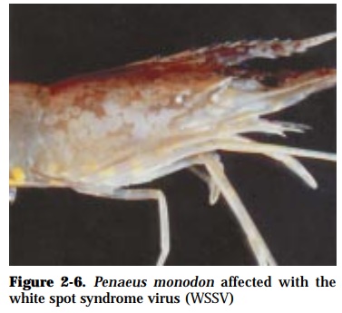
Typical signs of disease is the presence of distinct white cuticular spots (Fig. 2-6) (0.5-3 mm in diameter) most apparent at the exoskeleton and epidermis of diseased shrimp about 2 days after onset. The white spots start at the carapace and 5th and 6th abdominal segments that later affect the entire body shell. The moribund shrimp display red discoloration and have loose cuticle. Affected shrimps manifest sur-face swimming and gathering at pond dikes with broken antennae.

EFFECTS ON HOST:
This disease has been reported with the following names: White spot baculovirus (WSBV), White spot virus (WSV), Systemic ectodermal and mesodermal baculo-like virus (SEMBV), Chinese baculovirus (CBV), Hypodermal and hematopoietic necrosis baculo-like virus (HHNBV), Rod-shaped virus of Penaeus japonicus (RV-PJ), Penaeid acute viremia (PAV), Penaeid rod-shaped Dovavirus (PRDV).
Reduction in food consumption and empty gut develops followed by a rapid onset of the disease and high mortalities of up to 100% in 3 to 10 days. This disease affects a wide host range of crustaceans and targets various tissues (pleopods, gills, hemolymph, stom-ach, abdominal muscle, gonads, midgut, heart, periopods, lym-phoid organ, integument, nervous tissue and the hepatopan-creas) resulting in massive systemic pathology. Shrimps, 4-15 g, are particularly susceptible but the disease may occur from mysis to broodstock. Pre-moulting shrimps are usually affected. Penaeus indicus suffers earlier and greater losses compared to P. monodon. Crabs, krill and other shrimps are viral reservoirs. Pan-demic epizootics have occurred in extensive, semi-intensive and intensive culture systems regardless of water quality and salini-ties.
DIAGNOSIS:
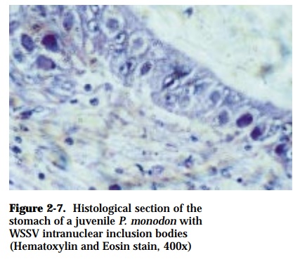
Clinical signs are diagnostic for this disease. However, recent reports indicate that some bacteria may induce similar signs, hence confirmation with other diagnostic tests should be done. Demonstration of the presence of hypertro-phied nuclei in stained squashes, smears of epithelial and connective tissues of the gills or stomach of affected shrimp. Histological sections show widespread cellular degeneration and severe nuclear hypertrophy, chromatin margination and eosinophilic intranuclear inclusions in the subcuticular epithelium of the shell, gill, stomach, connective tissues, hematopoietic tissues, lymphoid organ, antennal gland and nervous tissues (Fig. 2-7). Electron microscopy, PCR, DNA probe, Western Blot, and infection bioassay are confirmatony diagnostic tests.

Related Topics