Chapter: Medical Physiology: The Sense of Hearing
Transmission of Sound Waves in the Cochlea-ÔÇťTraveling WaveÔÇŁ
Transmission of Sound Waves in the CochleaÔÇöÔÇťTraveling WaveÔÇŁ
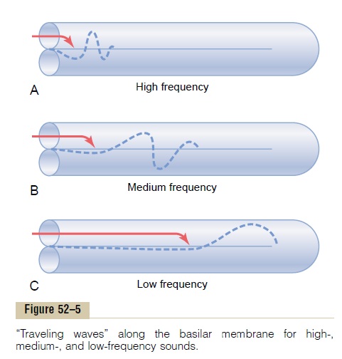
When the foot of the stapes moves inward against the oval window, the round window must bulge outwardbecause the cochlea is bounded on all sides by bony walls. The initial effect of a sound wave entering at the oval window is to cause the basilar membrane at the base of the cochlea to bend in the direction of the round window. However, the elastic tension that is built up in the basilar fibers as they bend toward the round window initiates a fluid wave that ÔÇťtravelsÔÇŁ along the basilar membrane toward the helicotrema, as shown in Figure 52ÔÇô5. Figure 52ÔÇô5A shows move-ment of a high-frequency wave down the basilar mem-brane; Figure 52ÔÇô5B, a medium-frequency wave; and Figure 52ÔÇô5C, a very low frequency wave. Movement of the wave along the basilar membrane is compara-ble to the movement of a pressure wave along the arte-rial walls; it is also comparable to a wave that travels along the surface of a pond.

Pattern of Vibration of the Basilar Membrane for Different Sound Frequencies. Note in Figure 52ÔÇô5 the differentpatterns of transmission for sound waves of different frequencies. Each wave is relatively weak at the outset but becomes strong when it reaches that portion of the basilar membrane that has a natural resonant fre-quency equal to the respective sound frequency. At this point, the basilar membrane can vibrate back and forth with such ease that the energy in the wave is dis-sipated. Consequently, the wave dies at this point and fails to travel the remaining distance along the basilar membrane. Thus, a high-frequency sound wave travels only a short distance along the basilar membrane before it reaches its resonant point and dies, a medium-frequency sound wave travels about halfway and then dies, and a very low frequency sound wave travels the entire distance along the membrane.
Another feature of the traveling wave is that it travels fast along the initial portion of the basilar membrane but becomes progressively slower as it goes farther into the cochlea. The cause of this is the high coefficient of elasticity of the basilar fibers near the oval window and a progressively decreasing coefficient farther along the membrane. This rapid initial trans-mission of the wave allows the high-frequency sounds to travel far enough into the cochlea to spread out and separate from one another on the basilar membrane. Without this, all the high-frequency waves would be bunched together within the first millimeter or so of the basilar membrane, and their frequencies could not be discriminated from one another.
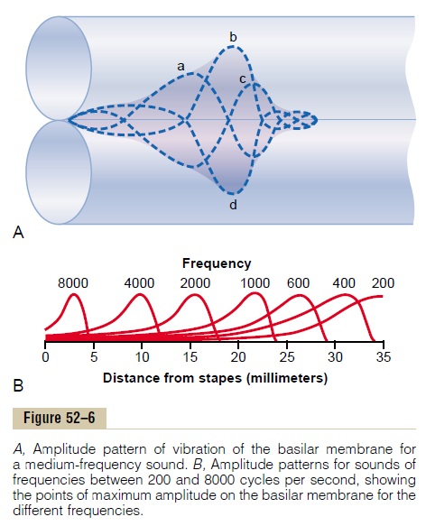
Amplitude Pattern of Vibration of the Basilar Mem- brane. The dashed curves of Figure 52ÔÇô6A show theposition of a sound wave on the basilar membrane when the stapes (a) is all the way inward, (b) has moved back to the neutral point, (c) is all the way outward, and (d) has moved back again to the neutral point but is moving inward. The shaded area around these different waves shows the extent of vibration of the basilar membrane during a complete vibratory cycle. This is the amplitude pattern of vibration of the basilar membrane for this particular sound frequency.

Figure 52ÔÇô6B shows the amplitude patterns of vibra-tion for different frequencies, demonstrating that the maximum amplitude for sound at 8000 cycles per second occurs near the base of the cochlea, whereas that for frequencies less than 200 cycles per second is all the way at the tip of the basilar membrane near the helicotrema, where the scala vestibuli opens into the scala tympani.
The principal method by which sound frequencies are discriminated from one another is based on the ÔÇťplaceÔÇŁ of maximum stimulation of the nerve fibers from the organ of Corti lying on the basilar membrane.
Related Topics