Chapter: Human Neuroanatomy(Fundamental and Clinical): Ventricles of the Brain
The Third Ventricle - Ventricles of the Brain
The Third Ventricle
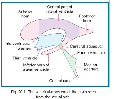
The third ventricle is the cavity of the diencephalon. It is a median cavity situated between the right and left thalami (Fig. 20.2). It communicates, on either side, with the lateral ventricle through the interventricular foramen (Figs. 20.1, 20.6). Posteriorly, it continues into the cerebral aqueduct which connects it to the fourth ventricle. The ventricle has two lateral walls, an anterior wall, a posterior wall, a floor and a roof.
Each lateral wall is marked by the hypothalamic sulcus (Fig. 20.6) which follows a curved course from the interventricular foramen to the aqueduct. Above the sulcus, the wall is formed by the medial surface of the thalamus. The two thalami are usually connected by a band of grey matter called the interthalamic connexus, which passes through the ventricle. The lateral wall, below the hypothalamic sulcus, is formed by the medial surface of the hypothalamus. A small part of the lateral wall, above and behind the thalamus, is formed by the epithalamus. The interventricular foramen is seen on the lateral wall, just behind the column of the fornix.
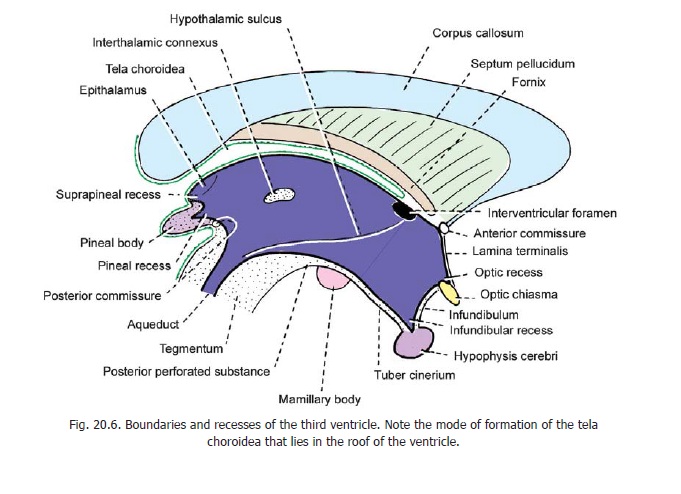
The anterior wall of the third ventricle is formed mainly by the lamina terminalis. Its upper part is formed by the anterior commissure, and by the columns of the fornix as they diverge from each other.
The posterior wall is formed by the pineal body and the posterior commissure.
The floor is formed by the optic chiasma, the tuber cinereum and the infundibulum, the mamillary bodies, the posterior perforated substance and the tegmentum of the midbrain.
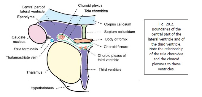
The roof of the ventricle is formed by the ependyma that stretches across the two thalami (Fig. 20.2). Above the ependyma there is the tela choroidea. Within the tela choroidea there are two plexuses of blood vessels (one on either side of the middle line) which bulge downwards into the cavity of the third ventricle. These are the choroid plexuses of the third ventricle (Fig. 20.7).
The cavity of the third ventricle shows a number of prolongations or recesses (Fig. 20.6). The infundibular recess extends into the infundibulum. The optic recess lies just above the opticchiasma. The pineal recess lies between the superior and inferior laminae of the stalk of the pineal body. The suprapineal recess lies above the pineal body in relationto the epithalamus.
Tela Choroidea of the third and lateral ventricles
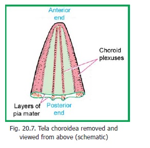
The tela choroidea is a double layered fold of pia mater that occupies the interval between the splenium of the corpus callosum and fornix, above, and the two thalami below. It is triangular in shape (Fig. 20.7). Its posterior end is broad and lies in the gap between the splenium (above) and the posterior part of the roof of the third ventricle (below) (Fig. 20.6). This gap is called the transverse fissure. The anterior end (representing theapex of the triangle) lies near the right and left interventricular foramina. The median part of the tela choroidea lies on the roof of the third ventricle. Its right and left lateral edges project into the central parts of the corresponding lateral ventricles (Fig. 20.2). When traced posteriorly, the two layers of pia mater forming the tela choroidea separate. The upper layer curves upwards over the posterior aspect of the splenium. The lower layer turns downwards over the pineal body and tectum (Fig. 20.6).
Choroid Plexuses
The choroid plexuses are highly vascular structures that are responsible for the formation of cerebrospinal fluid. The surface of each plexus is lined by a membrane formed by fusion of the ventricular ependyma with the pia mater of the tela choroidea. Deep to this membrane there is a plexus of blood vessels. Microscopic examination shows that the surface of the choroid plexus has numerous villous processes. Each process contains a plexus of capillaries that are connected to afferent and efferent vessels. Because of the presence of these processes the surface area of the choroid plexuses is considerable. It is further increased by the presence of microvilli (seen with the EM) present on the ependymal cells.
Four choroid plexuses are to be seen in relation to the tela choroidea of the third and lateral ventricles (Fig. 20.7). Two of these (one right and one left) lie along the corresponding lateral margins, and project into the central part of the corresponding lateral ventricle. Two other plexuses run parallel to each other, one on either side of the middle line. These are the choroid plexuses of the third ventricle. At each posterolateral angle of the tela choroidea the choroid plexus of the lateral ventricle continues into the inferior horn. The pial covering for this part of the plexus is provided by simple invagination of the pia, covering the medial aspect of the hemisphere, through the inferior part of the choroid fissure.
Related Topics