Mycetoma, Pathogenesis and Pathology, Classification of Mycetoma, Clinical Features, Laboratory Diagnosis | Medical Mycology - Subcutaneous Mycoses | 12th Microbiology : Chapter 9 : Medical Mycology
Chapter: 12th Microbiology : Chapter 9 : Medical Mycology
Subcutaneous Mycoses
Subcutaneous Mycoses
The
fungal infections are characterized by development of lesions at the site of
infection by the traumatic inoculation in the subcutaneous tissues. Examples
are Mycetoma, Sporotrichosis, Chromoblastomycosis and Rhinosporidiosis.
Mycetoma
Mycetoma
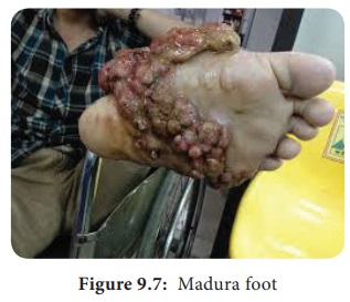
is a slowly progressive, chronic granulomatous infection of skin and
subcutaneous tissues with involvement of under lying fasciae and bones usually
affecting the extremities. Mycetoma is commonly called Madura foot or Maduramycosis
(Figure 9.7). They are
classified into two categories, namely eumycetoma
cased by fungi and actinomycetoma caused by higher
bacteria of class actinomycets.
Pathogenesis and Pathology
The
causative agent of Mycetoma is commonly present in saprobic soil source and is
transmitted by accidental trauma by thorns or by injury into the subcutaneous
tissue. It is common among farmers with minor trauma and abrasions of the skin.
Use of wicks for removal of earwax is responsible for Mycetoma of the ear.
HOTS: Is mycetoma occupational disease?
Classification of Mycetoma
Mycetoma
is classified on the basis of the causative agent. Aerobic actinomycetes causes actinomycetoma
whereas hyaline and phaeoid fungi cause
eumycetoma.
Clinical Features
The
clinical entity depends upon the age of the lesions and to size, shape and
color of the grains. The painless localized swollen lesions with purulent fluid
lead to the secondary bacterial infections. Important features of Mycetoma are
as follows:
i. Tumor like swelling.
ii. Multiple
draining sinuses.
iii. Presence of grains or granules in sinuses..
Laboratory Diagnosis
i. Samples
The clinical samples collected in Mycetoma is
usually grains, pus exudates or biopsy.
a. Direct Examination
Grams
staining, modified Ziehl – Neelson staining, LPCB and KOH wet mount are used to
visualize the organisms.
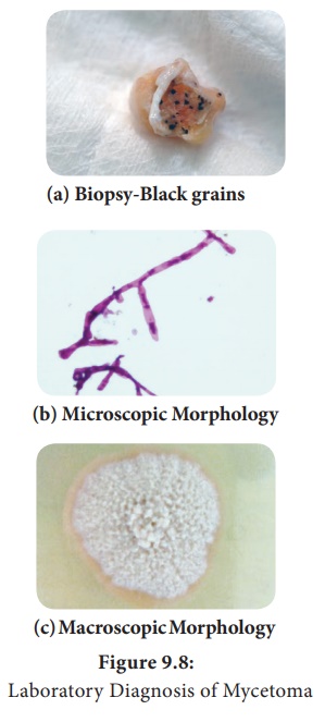
The grains should be washed, crushed and cultured
on different media. Crushed grains are examined (Figure 9.8a).
KOH mount
Eumycotic
grains show thick 2–6 µm hyphae with large globose swollen cells with or
without chlamydospores. Actinomycotic grains show thin filaments of 0.5–1 µm
with coccoid or bacillary forms.
Gram stain
Actinomycetoma
grains show Gram-positive branching filamentous bacteria with branches (Figure
9.8b).
Ziehl - Neelson stain
Nocardia species show red pink acid fast filamentous bacteria.
b. Culture
Crushed
grains are washed several times with normal saline without antibiotics and
inoculated on to Sabouraud dextrose agar, blood agar, Lowenstein -Jensen media
and brain-heart infusion agar. The plates are incubated at 25°C, 37°C and 44°C
for various organisms (Figure 9.8c).
ii. Treatment
1. Ketoconazole
200 mg and Itraconazole 100mg are given for 8–24 months to treat eumycetoma.
2. Sulfonamides, tetracylines, streptomy-cin, amoxicillin are administered to treat actinomycetoma.
Related Topics