Chapter: 11th Microbiology : Chapter 2 : Microscopy
Principles of Microscopy
Principles of Microscopy
All kind of microscopes use visible
light to observe specimens. Light has a number of properties that affect our
ability to visualise objects.
Properties of Light
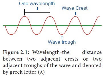
Light is a part of the wide spectrum
of electromagnetic radiation from the sun. It is a form of energy. The most
important property of light is wavelength (the length of light ray) (Figure
2.1).
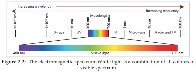
The sun produces a continuous
spectrum of electromagnetic radiation with waves of various lengths (Figure
2.2). Radiation of longer wavelength includes Infrared (IR) and radiowaves, the
shorter wavelengths include Ultra Violet (UV) rays and X-rays.
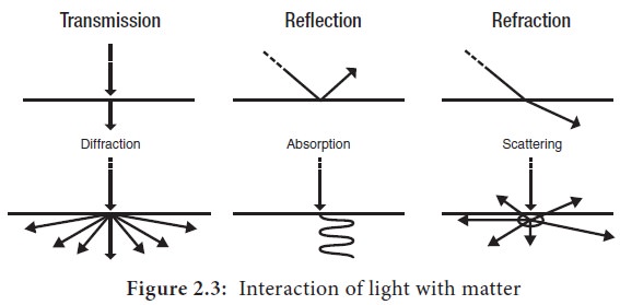
The physical behaviour of light can
be caterigorised as either light rays, light waves or light particles. The
combined properties of particle and wave enable light to interact with an
object in several different ways like transmission, absorption, reflection,
refraction, diffraction and scattering (Figure 2.3).
Lenses and its Properties
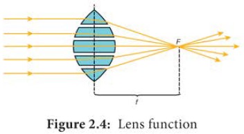
Lenses are optical devices which
focus or disperse a light beam by means of refraction. A simple lens consists
of a single piece of transparent material. Light rays from a distant source are
focused at the focal point F. The focal point lies at a distance f (focal length) from the lens’ centre (Figure 2.4).
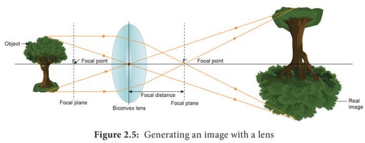
Generating an image with a lens
When an object is placed outside the
focal plane (the plane containing the focal point of the lens.), all the light
rays from the object are bent by the lens. The bent rays converge at the
opposite focal point. At the focal point, the light rays continue and converge
with nonparallel refracted light rays. The resultant reversed and magnified
image is formed in the plane of convergence (Figure 2.5).
Microscope resolution
Objective is the important part in
the microscope which is responsible to produce a clear image. The resolution of
the objective is most important. Resolution is the capacity of alens to
separate or distinguish between small objects that are close together. The
major factor in the resolution is the wave length of light used. The greatest
resolution obtained with light of the shortest wave length, that is the light
at the blue end of the visible spectrum are in the range of 450 to 500nm. The
highest resolution possible in compound light microscope is about 0.2μm. That means the two objects closer together than 0.2μm are not resolvable as distinct and separate. The light
microscope is equipped with three or four objectives. The working distance of
an objective is the distance between the front surface of the lens and the
surface of the cover glass or the specimen. Objectives with large numerical
apertures and great resolving power have short working distances.
Numerical aperture
Numerical aperture (NA) is the value representing the light gathering capacity of an objective lens. NA was first described by Ernst Abbe, and is defined by the following expression
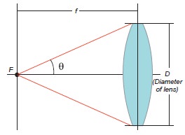
Numerical Aperture (NA) =n × sin(θ)
n = the refractive index of the medium between the specimen and objective; θ = half aperture angle or collection angle of the objective. (the maximum half angle of the cone of light that can enter or exit the lens).
The resolving power of a light
microscope depends on the wavelength of light used and the NA of the objective
lens.
The numerical aperture of a lens can
be increased by
·
Increasing the size of the lens
opening and/or
·
Increasing the refractive index of
the material between the lens and the specimen.
The larger the numerical aperture the
better the resolving power. It is important to illuminate the specimens
properly to have higher resolution. The concave mirror in the microscope
creates a narrow cone of light and has a small numerical aperture. However, the
resolution can be improved with a sub stage condenser. A wide cone of light
through the slide and into the objective lens increases the numerical aperture
there by improves the resolution of the microscope.
Types of microscopes
In order to view microorganism and
microbial structures of different sizes we require different kinds of
microscopes.
·
Light microscopes resolve images with
the help of light. The specimen is viewed as dark object against a light
background in bright field microscope. Dark field microscope uses a special
condenser and the specimen appears light against a black background. The other
types of mircoscopes are Phase contrast and Fluorescence microscope.
·
Electron microscope uses a beam of
electrons instead of light. Electrons pass through the specimen and form a two
dimensional image in Transmission Electron Microscope (TEM). Electrons are
reflected from the specimen and produce a three dimensional image in Scanning
Electron Microscope (SEM).
Related Topics