Chapter: 11th Microbiology : Chapter 2 : Microscopy
Bright Field Microscope
Bright Field Microscope
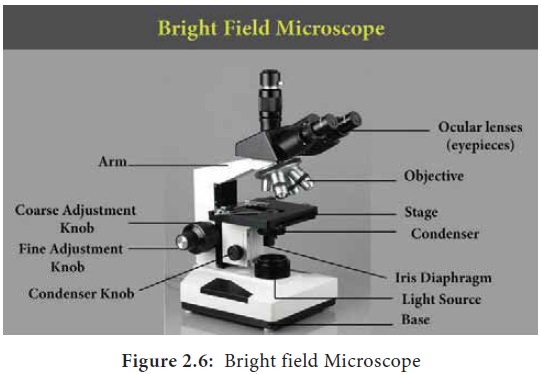
The most commonly used microscope for
general laboratory observations is the standard bright field microscope (Figure
2.6). It contains the following components
·
A mirror or an electric illuminator
is the light source which is located at the base of the microscope.

·
There are two focusing knobs, the
fine and the coarse adjustment knobs which are located on the arm. These are
used to move either the stage or the nosepiece to focus the image.
·
Mechanical stage is positioned about
half way up the arm, which allows precise contact on moving the slide.
·
The substage condenser is mounted
within or beneath the stage and focuses a cone of light on the slide. In the simpler
microscope, its position is fixed where as in advanced microscope it can be
adjusted vertically.
The upper part of microscope arm
holds the body assembly. The nose piece and one or more eyepieces or oculars
are attached to it. The body assembly contains series of mirrors and prisms so
that the barrel holding the eyepiece may be tilted for viewing. Three or five
objectives with different magnification power are fixed to the nosepiece and
can be rotated to the position beneath the body assembly. In bright field
microscopy; the specimen is viewed against a bright background. The details of
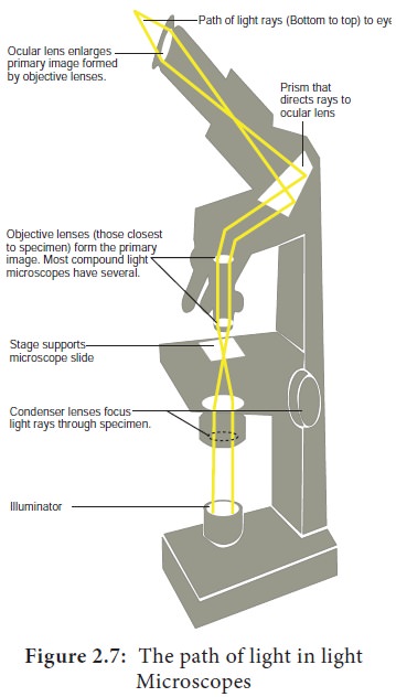
the image are defined by the surrounding light. A series of finely ground
lenses forms an image which is many times larger than the real image. This
magnification occurs when light rays from an illuminator (light source), pass
through a condenser which has lenses that direct the light rays through the
specimen. The light rays then pass into objective lens (the lens closest to the
specimen). The image is again magnified by the ocular lens or the eyepiece.
(Figure 2.7).

·
Magnification is the process of
enlarging the image of the specimen and can be calculated by multiplying the
objective lens magnification power by ocular lens magnification power.
Representative magnification values
for a 10X ocular are:
Scanning objective (4X) × (10X) = 40X
magnification
Low power objective (10X) × (10X) =
100X magnification
High dry objective (40X) × (10X) =
400X magnification
Oil immersion objective (100X) × (10X) = 1000X magnification
Oil Immersion
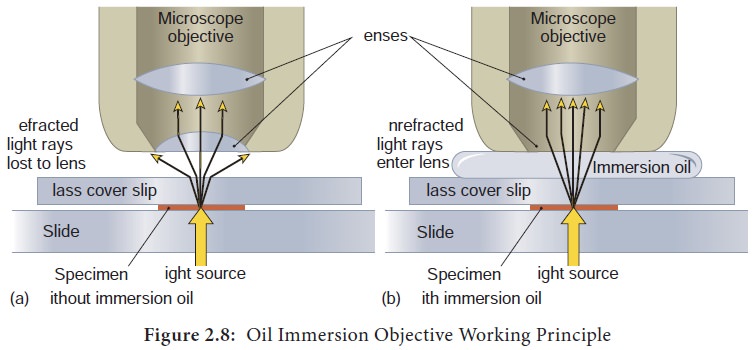
Oil immersion lens is designed to be
in direct contact with oil placed on the cover slip. An oil immersion lens has
a short focal length and hence there is a short working distance between the
objective lens and the specimen. Immersion oil has a refractive index closer to
that of glass than the refractive index of air, so the use of oil increases the
cone of light that enters the objective lens. Figure 2.8 explains the working
principle of oil immersion objective lens.

Related Topics