Chapter: Microbiology and Immunology: Bacteriology: Shigella
Pathogenesis and Immunity - Shigella
Pathogenesis and Immunity
◗ Virulence factors
Virulence in Shigella species involves both chromosomal- and plasmid-coded genes, which express for many virulence factors (Table 33-5).
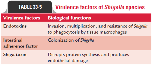
Endotoxins: The LPS moiety functions as an endotoxin and isan important component of the virulence of the bacteria. The endotoxin plays an important role in resistance of Shigella to nonspecific host defense encountered during tissue invasion. The toxin helps in invasion, multiplication, and resistance of Shigella to phagocytosis by tissue macrophages. The endotoxinincreases the cytotoxic activity of Shiga toxin on human vas-cular endothelial cells. The endotoxin is expressed by chromo-somal genes of the bacteria.
Intestinal adherence factor: Intestinal adherence factor isa 97-kDa outer membrane protein encoded by each gene on chromosomes. This mediates colonization of Shigella spp. in infected human hosts and in animal models.
Shiga toxin: Shiga toxin is an exotoxin produced byS. dysenteriae.It is a heat-labile protein and acts as enterotoxin and neurotoxin. Shiga toxin (Stx) is a group of cytotoxins that contain two major immunologically non–cross-reactive groups called Stx1 and Stx2. Both Stx1 and Stx2 groups are encoded by a bacteriophage inserted into the chromosome of the bacteria. Shiga toxins have one A subunit and five B subunits:
· The main function of B subunit is to bind toxins to host cell glycolipid (Gb3) surface receptor, present on the brush border of epithelial cell of the intestines. It also mediates transfer of the A subunit into the cell.
· Subunit A is a 32-kDa polypeptide. It cleaves the 28S rRNA in the 60S ribosomal subunit, thereby preventing the binding of aminoacyl-transfer RNA and disrupting protein synthesis.
The Shiga toxin shows three types of toxic activities:
a) Neurotoxic activity:This activity is demonstrable byparalysis and death of experimental animal following injection with the toxin. Although called neurotoxin, the primary site of its action is not the neural tissue but is the blood vessels, neurological manifestations being secondary.
b) Enterotoxic activity:These toxins are enterotoxic forligated rabbit intestinal segments with induction of fluid accumulation in ligated rabbit ileal loop. Two new Shigella enterotoxins, designated as S. ET-l and S. ET-2—the former confined to S. flexneri 2a and the latter more widespread— have been identified.
c) Cytotoxic activity:This is demonstrated by cytotoxicityof toxin for vero, HeLa, and some selected endothelial cells, such as human renal vascular endothelial cells. This appears to be the same as vero toxin 1 (or Shiga-like toxin) produced by certain strains ofEscherichia coli (VTEe).
The primary manifestation of Shiga toxin is damage to the intestinal epithelium of the infected host, causing diarrhea and dysentery. However, in a small number of patients, Shiga toxin can mediate damage to the glomerular endothelial cells, result-ing in hemolytic urinary syndrome.
◗ Pathogenesis of bacillary dysentery
Shigella spp. produce a serious disease known as bacillarydysentery. Infection occurs by ingestion. The infectivity dose (ID) is extremely low. As few as 10 S. dysenteriae bacilli can cause clinical disease, whereas 100–200 bacilli are needed for S. sonnei or S. flexneri infection.
Shigella spp. cause disease by invading and replicating in cells lin-ing the intestinal mucosa of the colon. Structural proteins, such as intestinal adhesion factor, endotoxin, and exotoxin mediate the adherence of the bacteria to the cells as well as their invasion, intra-cellular replication, and cell-to-cell spread. The bacilli infect the epithelial cells of the villi in the large intestine and multiply inside them. Subsequently, bacteria spread laterally to involve adjacent cells and penetrate into the lamina propria (Fig. 33-1). Shigellae lyse the phagocytic vacuole and multiply in the host cell cyto-plasm. Shigellae survive phagocytosis by inducing programed cell death or apoptosis. This mechanism also leads to the release of interleukin-1b resulting in the attraction of polymorphonu-clear leukocytes into the infected tissues. This in turn alters the integrity of the intestinal wall and allows the bacteria to reach the deeper epithelial cells. Shiga toxin produced by the bacteria also plays an important role in progression of mucosal lesions after invasion of the colonic cells. The toxin also induces vascu-lar damage in the colonic mucosa.

Mucosal edema, erythema, friability, superficial ulceration, and focal mucosal hemorrhage involving the rectosigmoid junction are the typical pathological features primarily observed in the condition.
Related Topics