Chapter: Clinical Cases in Anesthesia : Thrombocytopenia In Pregnancy
Describe the thromboelastogram and its limitations
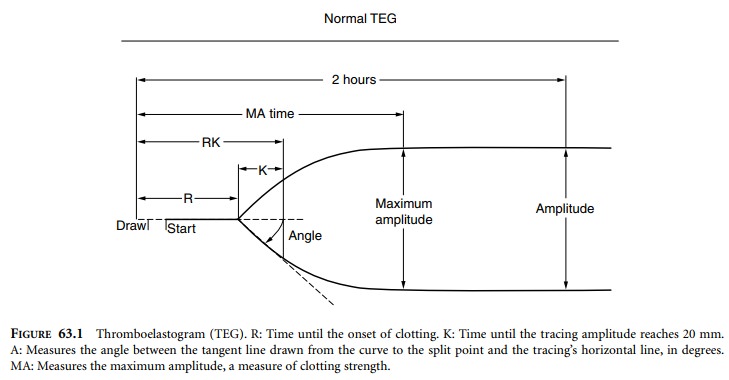
Describe the thromboelastogram and its limitations.
The thromboelastogram (TEG) measures all phases
of coagulation and fibrinolysis by using less than 1 mL of a whole blood sample
to measure the shear elasticity of clot-ting blood. Blood is placed in a
cylindrical cup that oscillates. A pin is suspended in the blood by a torsion
wire and is monitored for motion. The torque of the rotating cup only affects
the pin after fibrin–platelet bonding has linked the cup and pin together. The
strength of the developing clot affects the magnitude of the pin motion such
that strong clots move the pin directly in phase with the cup and weak clots do
not. The resulting profile is a measure of the time it takes for the first
fibrin strand to form, the kinetics of the clot, strength of the clot, and
breakdown of the clot (Figure 63.1). The maximum amplitude (MA) has been found
to correlate best with platelet function.
Orlikowski et al. (1996) measured platelet
counts, TEG parameters, and bleeding times in healthy pregnant women and in
those with preeclampsia. They found that the MA remains normal (53 mm) until the
platelet count decreases to less than 54,000 mm−3 (95%
confidence limit 40,000–75,000 mm−3). Based on their study, they suggested that a platelet count of
75,000 mm−3 should
be associated with adequate hemostasis. There is no clinical evidence, however,
that a normal MA correlates with safe epidural anesthesia.
Related Topics