Chapter: Clinical Cases in Anesthesia : Depolarizing Neuromuscular Blockade
Describe the anatomy and physiology of normal neuromuscular transmission
Describe
the anatomy and physiology of normal neuromuscular transmission.
Neuromuscular transmission begins with
depolariza-tion down the axon of a motor nerve to the motor nerve terminal (Figure
23.1). The wave of depolarization is prop-agated by sequential opening of
sodium channels. This allows sodium to enter the cell, increasing the
intracellular concentration of positively charged ions. The transmem-brane
potential is altered from negative to positive in the area of the open sodium
channel. This electrical event causes the next sodium channel to open and
continues the process until the nerve terminal is reached. When the nerve
terminal is thus depolarized, calcium channels open, allow-ing calcium to
enter. The rise in intracellular calcium facil-itates the binding of
intracellular “packets” or quanta of acetylcholine to the cell wall bordering
the neuromuscular junction. Acetylcholine molecules are then released into the
junctional (synaptic) cleft.
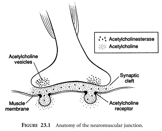
Nicotinic acetylcholine receptors on the motor
endplate of the muscle cell have binding sites for acetylcholine. This receptor
is made up of five transmembrane protein chains, two of which are identical and
contain one acetylcholine receptor site each. When two acetylcholine molecules
bind to these two sites, the receptor is activated to respond as an ion channel
for the rapid ingress of sodium. This move-ment of positively charged ions into
the cell creates a small electrical current. When enough receptors are
activated, the summation of these small electrical currents becomes sufficient
to depolarize the endplate.
Depolarization of the endplate stimulates the
sodium channels in the perijunctional area to open, and thus the electrical
current is propagated along the myocytes by sequential opening of sodium
channels. When the myocytes are depolarized, stored calcium ions from the
sarcoplasmic reticulum are released intracellularly. The free intracellular
calcium ions activate myosin adenosine triphosphatase (ATPase), which
precipitates excitation– contraction coupling of actin and myosin proteins to
cause contraction.
At the same time, acetylcholine, which
activated the receptors to initiate this process, is rapidly metabolized by
acetylcholinesterase. Acetylcholinesterase is a protein on the muscle membrane
in the junctional cleft that rapidly causes the hydrolysis of acetylcholine.
Acetylcholine may alterna-tively be actively taken up into the nerve terminal
from which it was released. This removal of acetylcholine from the
neuromuscular junction allows for repolarization of the motor endplate.
Subsequently, sodium channels along the muscle cell close, and the muscle cell
repolarizes. Calcium is actively taken up into the sarcoplasmic reticulum and
the actin-myosin fibers reset, allowing the muscle to relax.
Related Topics