Chapter: Pathology: Cardiac Pathology
Congenital Heart Disease

CONGENITAL HEART DISEASE
Congenital
heart disease is the most common cause of childhood heart disease inthe
United States; 90% of cases are idiopathic and 5% are associated with genetic
disease (trisomies, cri du chat, Turner syndrome, etc.), viral infection
(especially congenital rubella), or drugs and alcohol.
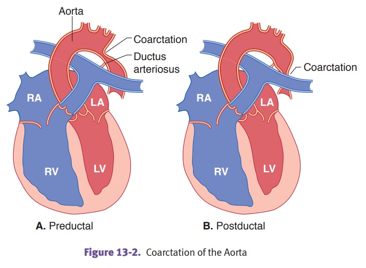
Coarctation
of the aorta is a segmental narrowing of the aorta.
·
Preductal coarctation (infantile-type)
is associated with Turner syndrome andcauses severe narrowing of aorta proximal
to the ductus arteriosus. It is usu-ally associated with a patent ductus
arteriosus (PDA), which supplies blood to aorta distal to the narrowing, and
right ventricular hypertrophy (secondary to the need for the right ventricle to
supply the aorta through the patent ductus arteriosus). It presents in infancy
with congestive heart failure that is accom-panied by weak pulses and cyanosis
in the lower extremities; the prognosis is poor without surgical correction.
·
Postductal coarctation (adult-type)
causes stricture or narrowing of the aortadistal to the ductus arteriosus. It
can present in a child or an adult with hyper-tension in the upper extremities,
and hypotension and weak pulses in the lower extremities. Some collateral
circulation may be supplied via the internal mammary and intercostal arteries;
the effects of this collateral circulation may be visible on chest x-ray with
notching of the ribs due to bone remodeling as a consequence of increased blood
flow through the intercostal arteries.
Complications
can include congestive heart failure (the heart is trying too hard),
intracerebral hemorrhage (the blood pressure in the carotid arteries is too
high), and dissecting aortic aneurysm (the blood pressure in the aortic route
is too high).

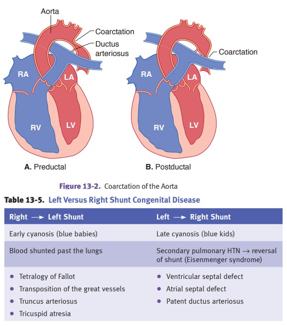
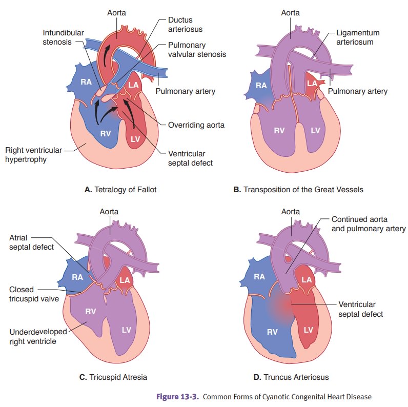
Tetralogy of Fallot is
the most common cause of congenital cyanotic heart disease.The classic tetrad
includes right ventricular outflow
obstruction/stenosis; rightventricular hypertrophy; ventricular septal defect; and overriding aorta. Clinicalfindings
include cyanosis, shortness of breath, digital clubbing, and polycythemia.
Progressive pulmonary outflow stenosis and cyanosis develop over time;
treatment is surgical correction.

Transposition of the great vessels is
an abnormal development of the truncoconalseptum whereby the aorta arises from
the right ventricle, and the pulmonary artery arises from the left ventricle.
The risk is increased in infants of diabetic mothers. Affected babies develop
early cyanosis and right ventricular hypertrophy. To survive, infants must have
mixing of blood by a VSD, ASD, or PDA. The prognosis is poor without surgery.

Truncus
arteriosus is a failure to develop a dividing septum between the aorta
andpulmonary artery, resulting in a common trunk. Blood flows from the
pulmonary trunk to the aorta. Truncus arteriosus causes early cyanosis and
congestive heart failure, with a poor prognosis without surgery.
Tricuspid
atresia refers to the absence of a communication between the right
atriumand ventricle due to developmental failure to form the tricuspid valve.
Associated defects include right ventricular hypoplasia and an ASD. The
prognosis is poor without surgery.
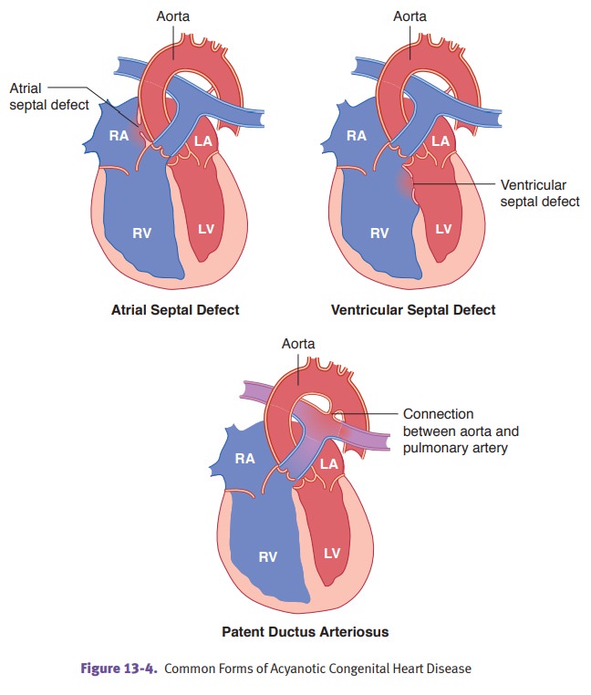
Ventricular
septal defect (VSD), which consists of a direct
communication betweenthe ventricular chambers, is the second most common
congenital heart defect (the most common is a bicuspid aortic valve).
·
A small ventricular septal defect may be asymptomatic and close
sponta-neously, or it may produce a jet stream that damages the endocardium and
increases the risk of infective endocarditis.
·
A large ventricular septal defect may cause Eisenmenger complex,
which is characterized by secondary pulmonary hypertension, right ventricular
hyper-trophy, reversal of the shunt, and late cyanosis.
·
In both types, a systolic murmur can be heard on auscultation.
Ventricular septal defects are commonly associated with other heart defects.
Large ven-tricular septal defects can be surgically corrected.
Atrial
septal defect (ASD) is a direct communication between
the atrial chambers.
The
most common type is an ostium secundum defect. Complications include
Eisenmenger syndrome and paradoxical emboli.
Patent
ductus arteriosus (PDA) is a direct communication
between the aorta andpulmonary artery due to the continued patency of the
ductus arteriosus after birth. It is associated with prematurity and congenital
rubella infections. Clinical findings include machinery murmur, late cyanosis,
and congestive heart failure. Eisenmenger syndrome may develop as a
complication.
Related Topics