Chapter: Human Nervous System and Sensory Organs : Brain's Cerebrovascular and Ventricular Systems
Arteries - Cerebrovascular Systems
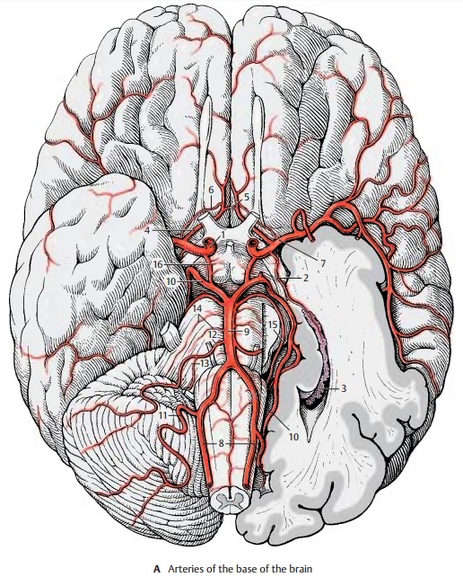
Cerebrovascular System
Arteries
The brain is supplied by four large arteries: the two internal carotid arteries and the two vertebral arteries.
The internal carotid artery (A1) passes through the dura mater medially to the anterior clinoid process of the sphenoid bone. Between the subarachnoid and the pia mater, it gives off the superior hypophysialartery, the ophthalmic artery,the posterior communicating artery (A16), and the anterior choroidal artery (A2). It then divides into two large terminal branches, the anterior cerebral artery (A4) and the middle cerebral artery (A7).
The anterior choroidal artery (A2) runs along the optic tract to the choroid plexus (A3) in the inferior horn of the lateral ventricle. It gives off fine branches that supply the optic tract, the temporal genu of the optic radia-tion, the hippocampus, the tail of the cau-date nucleus, and the amygdaloid body.
The anterior cerebral artery (A4) runs on the medial surface of the hemisphere across the corpus callosum. The two anterior cerebral arteries are interconnected by the anteriorcommunicating artery (A5). The long centralartery (recurrent artery of Heubner) (A6) branches off shortly after the communicat-ing artery. It passes through the anterior perforated substance into the brain and supplies the anterior limb of the internal capsule, the adjacent region of the head of the caudate nucleus, and the putamen.
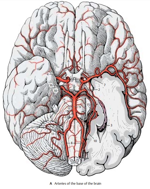
The middle cerebral artery (A7) runs laterally toward the lateral sulcus; above the perforated substance it gives off 8 – 10 striatebranches that enter the brain. At the en-trance of the lateral fossa, it divides into several large branches that spread over the lateral surface of the hemisphere.
The two vertebral arteries (A8) arise from the two subclavian arteries and enter the cranial cavity through the foramen mag-num; they unite at the upper margin of the medulla oblongata to form the unpairedbasilar artery (A9). The latter ascends alongthe ventral surface of the pons and bifur-cates at the upper margin of the pons into the two posterior cerebral arteries (A10). The vertebral artery gives off the posterior infe-rior cerebellar artery (A11), which suppliesthe lower surface of the cerebellum and the choroid plexus of the fourth ventricle. The basilar artery gives off the anterior inferiorcerebellar artery (A12), which also suppliesthe lower surface of the cerebellum as well as the lateral parts of medulla and pons. The labyrinthine artery (A13) runs as a finebranch together with the facial nerve and the vestibulocochlear nerve through the in-ternal acoustic meatus into the inner ear. It may arise from the basilar artery or from the anterior inferior cerebellar artery. Numer-ous small branches reach as pontine arteries (A14) directly into the pons. The superiorcerebellar artery (A15) runs along the uppermargin of the pons and extends deep into the cisterna ambiens around the cerebral peduncles to the dorsal surface of the cerebellum.
The circle of Willis.Theposterior communi-cating arteries (A16) connect on both sidesthe posterior cerebral arteries with the in-ternal carotid arteries so that the blood flow of the vertebral arteries can communicate with the carotid circulation. The anterior cerebral arteries are interconnected through the anterior communicating artery. This way, a closed cerebral arterial circle is created at the base of the brain. However, the anastomoses are often so thin that they do not allow for a significant exchange of blood. Under conditions of normal in-tracranial pressure, each hemisphere is fed by the ipsilateral internal carotid artery and the ipsilateral posterior cerebral artery.
Internal Carotid Artery (A – C)
The internal carotid artery (C1) can be sub- divided into a cervical part (between the carotid bifurcation and the base of the skull); a petrosal part (in the carotid canal ofthe petrous bone); a cavernous part ; and a cerebral part. The cavernous and cerebral seg- ments of the artery form an S-shaped curve (carotid siphon) (C2). The inferior hypophysial artery comes off in the cavernous part, followed by small branches to the dura and to cranial nerves IV and V. After giving off the superior hypophysial artery , the ophthalmic artery, and the anterior choroidal artery in the cere- bral part, the internal carotid artery dividesinto two large terminal branches, the ante- rior cerebral artery and the middle cerebral artery.
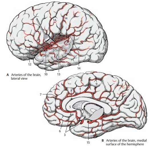
The anterior cerebral artery (BC3) turns toward the longitudinal cerebral fissureafter giving off the anterior communicating artery. The postcommunical part of theartery (pericallosal artery) (BC4) runs on the medial hemispheric wall around the ros-trum and the genu of the corpus callosum (B5) and along the superior surface of the corpus callosum toward the parieto-occipi- tal sulcus. It gives off branches to the basal surface of the frontal lobe (medial fron- tobasal artery) (B6). The other branches spread over the medial surface of the hemi- sphere; they are the frontal branches (BC7), the callosomarginal artery (BC8), and the paracentral artery (B9) which supplies the motor area for the leg.
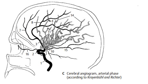
The middle cerebral artery (AC10) extends laterally to the bottom of the lateral fossa, where it divides into several groups of branches. The artery is divided into three segments. The sphenoidal part gives off the central arteries (fine branches for the stri- atum, thalamus, and internal capsule), and the insular part gives rise to the short insular arteries (C11) for the insular cortex, the lateral frontobasal artery (A12), and the tem- poral arteries (A13) for the cortex of the temporal lobe. The last segment, the termi- nal part, is formed by the long branches for the cortex of the central region and the parietal lobe (AC14). There are considerable variations regarding the bifurcation and course of individual arteries.
The posterior cerebral artery (BC15) develops as a branch of the internal carotid artery. In adults, however, it lies relatively far caudally. It is only connected with the inter- nal carotid artery through the thin posterior communicating artery. Because most of its blood comes from the vertebral arteries, it is regarded as part of their supply area; the latter consists of the subtentorial parts of the brain (brain stem and cerebellum) and the supratentorially located occipital lobe, the basal part of the temporal lobe, and the caudal parts of the striatum and thalamus (tentorium of the cerebellum, p. 288, B5). All parts of the frontal brain lying in front of it receive their blood from the internal carotid artery.
The posterior cerebral artery ramifies on the medial surface of the occipital lobe and on the basal surface of the temporal lobe. It gives off the posterior choroidal artery for the choroid plexus of the third ventricle as well as fine branches for the striatum and thalamus. The ophthalmic artery (C16) runs from the internal carotid artery to the orbit. Carotid angiogram (arterial phase). A dia- gram of a carotid angiogram is shown in C. For diagnostic purposes, a contrast medium is injected into the internal carotid artery. Within a few seconds, the contrast medium spreads through the supply area of the artery. A radiograph taken immediately il- lustrates the arterial vascular tree. Whenviewing arteriographic images, it must be taken into consideration that all blood ves- sels are seen in a single plane (see contrast radiography, p. 264, A).
Areas of Blood Supply
Anterior Cerebral Artery (A, B)
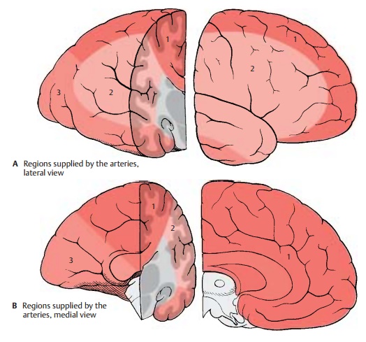
The short branches of the anterior cerebralartery (AB1) extend to the optic chiasm, tothe septum pellucidum, to the rostrum and genu of the corpus callosum, while the long recurrent artery of Heubner (long centralartery) runs to the medial part of the head of the caudate nucleus and to the anterior limb of the internal capsule. The cortical branches supply the medial parts of the base of the frontal brain and the olfactory lobe, and also the frontal and parietal cortex on the medial surface of the hemisphere and the corpus callosum as far as the splenium. The supply area of the artery ex-tends beyond the edge of the pallium to the dorsal convolutions of the convexity.
Middle Cerebral Artery (A, B)
The striate branches of the middle cerebralartery (AB2) terminate in the globus pal-lidus, in parts of the thalamus, in the genu and parts of the anterior limb of the internal capsule. The branches given off by the insu-lar arteries ramify in the insular cortex and in the claustrum and reach into the external capsule. The area supplied by the cortical branches includes the lateral surface of frontal, parietal, and temporal lobes and also a large part of the central region and the temporal pole. The branches not only supply the cortex but also the white matter as far as the lateral ventricle, including the central part of the optic radiation.
Posterior Cerebral Artery (A, B)
The posterior cerebral artery (AB3) gives off fine short branches that supply the cerebral peduncles, pulvinar, geniculate bodies, quadrigeminal plate, and splenium of the corpus callosum. The cortical supply area occupies the basal part of the temporal lobe and the occipital lobe with the visual cortex (striate area); however, the latter is also reached in the region of the occipital pole by the most inferior branches of the middle cerebral artery.
Blood Supply for the Nuclei of Diencephalon and Telencephalon (C, D)
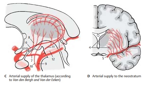
The recurrent artery of Heubner and the striate branches (D4) of the middle cerebralartery (D5) supply the head of the caudate nucleus, putamen, and internal capsule. The role of the anterior choroidal artery (C6) in the supply of deep structures varies; its branches extend not only to the hippocam-pus and amygdaloid body but also to parts of the pallidum and thalamus. The rostral part of the thalamus receives a branch from the posterior communicating artery (C7), the thalamic branch (C8). The middle and caudal parts of the thalamus are supplied by the basilar artery (C9), from where direct branches (C10) may run to the thalamus. Other fine thalamic branches are given off by the posterior choroidal artery (C11) and the posterior cerebral artery (C12).
Vascularization.The large cerebral vesselslie without exception on the surface of the brain. They give off small arteries and arteri-oles that penetrate vertically into the brain and ramify. The capillary network is very dense in the gray matter but far less so in the white matter.
Clinical Note: Sudden obstruction of an arteryby a thrombus, air bubble, or fat droplet in the bloodstream (embolism) causes the death of brain tissue in the supply area of the affected artery. The anastomoses between the vascular areas are not adequate to supply the affected region through nearby areas in case of a sudden obstruc-tion. Especially affected are the middle cerebralartery and its branches.
Related Topics