Chapter: Human Nervous System and Sensory Organs : Brain's Cerebrovascular and Ventricular Systems
Internal Carotid Artery - Cerebrovascular Systems

Internal Carotid Artery
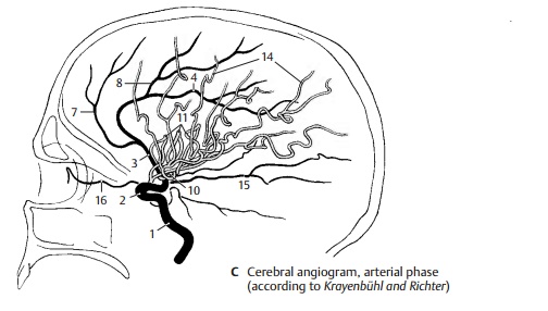
The internal carotid artery (C1) can be sub- divided into a cervical part (between the carotid bifurcation and the base of the skull); a petrosal part (in the carotid canal ofthe petrous bone); a cavernous part; and a cerebral part. The cavernous and cerebral seg- ments of the artery form an S-shaped curve (carotid siphon) (C2). The inferior hypophysial artery comes off in the cavernous part, followed by small branches to the dura and to cranial nerves IV and V. After giving off the superior hypophysial artery , the ophthalmic artery, and the anterior choroidal artery in the cere- bral part, the internal carotid artery dividesinto two large terminal branches, the ante- rior cerebral artery and the middle cerebral artery.

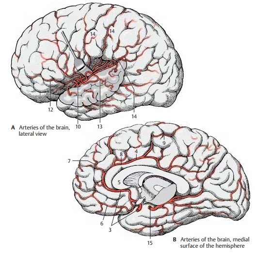
The anterior cerebral artery (BC3) turns toward the longitudinal cerebral fissureafter giving off the anterior communicating artery. The postcommunical part of theartery (pericallosal artery) (BC4) runs on the medial hemispheric wall around the ros-trum and the genu of the corpus callosum (B5) and along the superior surface of the corpus callosum toward the parieto-occipi- tal sulcus. It gives off branches to the basal surface of the frontal lobe (medial fron- tobasal artery) (B6). The other branches spread over the medial surface of the hemi- sphere; they are the frontal branches (BC7), the callosomarginal artery (BC8), and the paracentral artery (B9) which supplies the motor area for the leg.

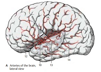
The middle cerebral artery (AC10) extends laterally to the bottom of the lateral fossa, where it divides into several groups of branches. The artery is divided into three segments. The sphenoidal part gives off the central arteries (fine branches for the stri- atum, thalamus, and internal capsule), and the insular part gives rise to the short insular arteries (C11) for the insular cortex, the lateral frontobasal artery (A12), and the tem- poral arteries (A13) for the cortex of the temporal lobe. The last segment, the termi- nal part, is formed by the long branches for the cortex of the central region and the parietal lobe (AC14). There are considerable variations regarding the bifurcation and course of individual arteries.
The posterior cerebral artery (BC15) develops as a branch of the internal carotid artery. In adults, however, it lies relatively far caudally. It is only connected with the inter- nal carotid artery through the thin posterior communicating artery. Because most of its blood comes from the vertebral arteries, it is regarded as part of their supply area; the latter consists of the subtentorial parts of the brain (brain stem and cerebellum) and the supratentorially located occipital lobe, the basal part of the temporal lobe, and the caudal parts of the striatum and thalamus (tentorium of the cerebellum, p. 288, B5). All parts of the frontal brain lying in front of it receive their blood from the internal carotid artery.
The posterior cerebral artery ramifies on the medial surface of the occipital lobe and on the basal surface of the temporal lobe. It gives off the posterior choroidal artery for the choroid plexus of the third ventricle as well as fine branches for the striatum and thalamus. The ophthalmic artery (C16) runs from the internal carotid artery to the orbit. Carotid angiogram (arterial phase). A dia- gram of a carotid angiogram is shown in C. For diagnostic purposes, a contrast medium is injected into the internal carotid artery. Within a few seconds, the contrast medium spreads through the supply area of the artery. A radiograph taken immediately il- lustrates the arterial vascular tree. Whenviewing arteriographic images, it must be taken into consideration that all blood ves- sels are seen in a single plane (see contrast radiography, p. 264, A).
Related Topics