Chapter: Pathology: Red Blood Cell Pathology: Anemias
Anemias
ANEMIAS
Anemia is
a reduction below normal limits of the total circulating red cell mass. Signsof
anemia include palpitations, dizziness, angina, pallor of skin and nails,
weakness, claudication, fatigue, and lethargy.
·
Reticulocytes
are immature, larger red cells (macrocytic cells) that are
spheri-cal and have a bluish color (polychromasia) due to free ribosomal RNA.
Reticulocytes do not have a nucleus; note that any erythrocyte with a nucleus
(nRBC) in peripheral blood is abnormal. Reticulocyte maturation to a mature
erythrocyte takes about 1 day. The reticulocyte count is the percentage of red
immature cells present in peripheral blood (normal 0.5–1.5%).
The corrected reticulocyte count takes into consideration
the degree of anemia and is calculated as (patient’s hct/45) × (reticulocyte
count); the idea behind the calculation is to scale the reticulocyte count by
multiplying by the ratio of the patient’s hematocrit to “normal” hematocrit of
45%. When interpreting the corrected reticulocyte count, <2% indicates poor
bone marrow response and >3% indicates good bone marrow response.
·
The reticulocyte production index is
the corrected reticulocyte count/2; use this measure if bone marrow
reticulocytes (shift cells) are present (polychro-masia). The division by 2 is
because shift cells take twice as long as reticulo-cytes to mature (2 days
versus 1 day).
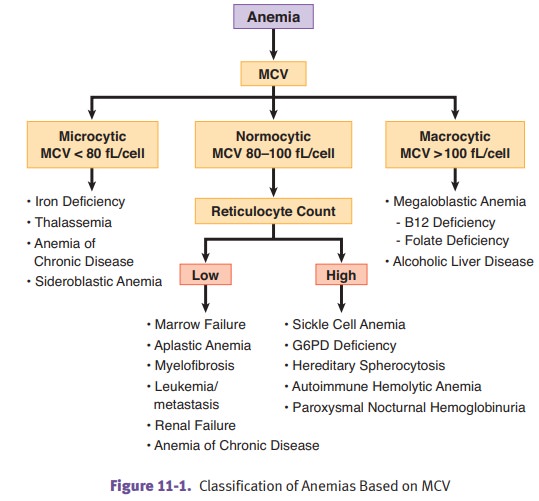
Classification
of anemia can be based on color: normochromic anemias have nor-mal red cell
color (central pallor of about a third the diameter of the erythrocyte);
hypochromic anemias have decreased color (seen as an increased central pallor
of erythrocyte); and hyperchromic anemias, while theoretically possible, are
usually instead called spherocytosis and have increased color (loss of central
pallor of eryth-rocyte). Classification of anemia can also be based on size
(MCV).
The
pathogenesis of anemia varies with the underlying disease. Blood loss can cause
anemia. Hemolytic anemias are also
important, and include hereditary spherocyto-sis, glucose-6-phosphate
dehydrogenase deficiency, sickle cell disease, hemoglobin C disease,
thalassemia, and paroxysmal nocturnal hemoglobinuria. Immunohe-molytic anemias, which are hemolytic anemias with an
immune component to thepathology, include autoimmune hemolytic anemia (AIHA),
cold AIHA, incompat-ible blood transfusions, and hemolytic disease of the
newborn. Anemias of dimin-ished
erythropoiesis include megaloblastic anemia (B12 and folate
deficiencies),iron deficiency anemia, anemia of chronic disease, aplastic
anemia, myelophthisic anemia, and sideroblastic anemia.
Related Topics