Chapter: Human Neuroanatomy(Fundamental and Clinical): The Spinal Cord : Gross Anatomy and Some features of Internal Structure
The Spinal Cord : Gross Anatomy and Some features of Internal Structure
The Spinal Cord : Gross Anatomy and Some features of Internal Structure
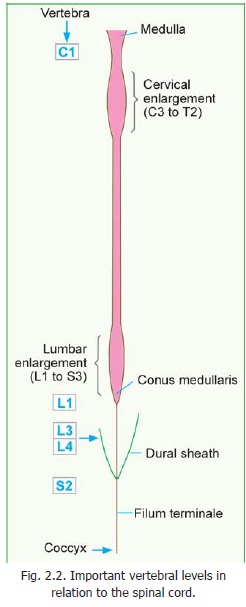
The spinal cord (or spinal medulla) is the most important content of the vertebral canal. The upper end of the spinal cord becomes continuous with the medulla oblongata at the level of the upperborder of the first cervical vertebra. The lower end of the spinal cord lies at the level of the lowerborder of the first lumbar vertebra. The level is, however, variable and the cord may terminate one vertebra higher or lower than this level. The level also varies with flexion or extension of the spine.
The lowest part of the spinal cord is conical and is called the conus medullaris. The conus is continuous, below, with a fibrous cord called the filum terminale (Fig. 2.2).
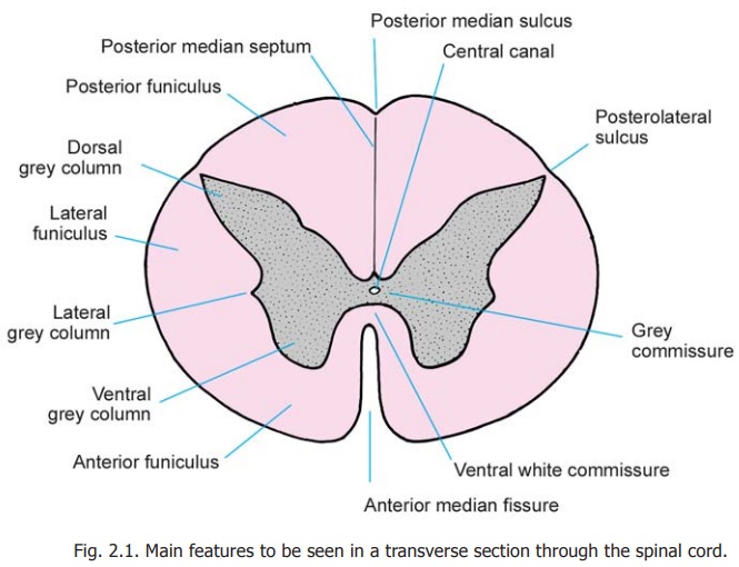
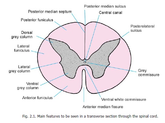
When seen in transverse section the grey matter of the spinal cord forms an H-shaped mass (Fig. 2.1). In each half of the cord the grey matter is divisible into a larger ventral mass, the anterior (orventral) grey column, and a narrow elongated posterior (or dorsal) grey column. In someparts of the spinal cord a small lateral projection of grey matter is seen between the ventral and dorsal grey columns. This thelateral grey column. The grey matter of the right and left halves of the spinal cord is connected across the middle line by the grey commissure which is traversed by the central canal. The lower end of the central canal expands to form the terminal ventricle which lies in the conus medullaris. The cavity within the spinal cord continues for a short distance into the filum terminale. The central canal of the spinal cord contains cerebrospinal fluid. The canal is lined by ependyma.
The white matter of the spinal cord is divided into right and left halves, in front by a deep anteriormedian fissure, and behind by the posterior median septum. In each half of the cord the whitematter medial to the dorsal grey column forms the posterior funiculus (or posterior white column).
The white matter medial and ventral to the anterior grey column forms the anterior funiculus (or anterior white column), while the white matter lateral to the anterior and posterior grey columns forms the lateral funiculus. (The anterior and lateral funiculi are collectively referred to as the anterolateral funiculus).
The white matter of the right and left halves of the spinal cord is continuous across the middle line through the ventralwhite commissure which lies anterior to the grey commissure. Some myelinated fibres running transverselyin the grey commissure, posterior to the central canal are referred to as the dorsal white commissure.
Related Topics