Chapter: Health Management in Aquaculture: Viral diseases
Monodon Baculovirus (MBV) Disease - Viral Infections in Penaeid Shrimps
Monodon Baculovirus (MBV) Disease
CAUSATIVE AGENT:
P. monodon-type baculovirus (75 x 300 nm)
SPECIES AFFECTED:
The giant tiger prawn Penaeus monodon, and other penaeid shrimps like P.merguiensis, P. vannamei, P. esculentus, P. semisulcatus, P. penicillatus, P. plebejus, P. kerathurus.
GROSS SIGNS:
Affected shrimps exhibit pale-bluish-gray to dark blue-black coloration, slug-gish and inactive swimming movements, loss of appetite and retarded growth. An increased growth of benthic diatoms and filamentous bacteria may cause fouling on the exoskeleton/gills. Infected pond-reared shrimps at 45 days of culture (DOC) stocked at 4 to 100 per m2 manifested slow growth rates and pale yellow to reddish brown hepatopancreas.
EEFFECT ON HOST:
This is among the first viral infections diagnosed in mysis, postlarvae, juveniles and adults of the giant tiger prawn, Penaeus monodon. The virus causes de-struction of the hepatopancreas and lining of the digestive tract. Spherical, eosinophilic occlusion bodies fill up enlarged nuclei of hepatopancreatic cells and are discharged into the lumen after cells have been destroyed. This may be followed by necrosis with secondary bacterial infection. PL-3 is the earliest stage found infected with MBV. However, experimental waterborne inoculation of MBV to mysis-2 (M-2), postlarvae-3 (PL-3), PL-6, PL-9 and PL-11 resulted in MBV infections within 12 days post-inoculation. The incidence rate of MBV was reported at 20-100%. Cumulative mortality of 70% was observed amongP.monodon juveniles cultured in raceways and tanks. It is associated with a highincidence of bacterial infections expressed as localized “shell disease.” In addi-tion, significant mortalities can occur during stress and crowding.
DIAGNOSIS:
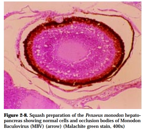
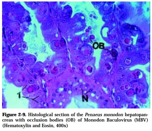
Demonstration of occlusion bodies in wet mounts of feces, midgut or hepato-pancreas stained with malachite green (Fig. 2-8). Histological sections show the presence of eosinophilic, multiple occlusion bodies within the hypertro-phied nuclei of the hepatopancreatic tissues with the following development (Fig. 2-9). Other diagnostic tests are DNA probe and PCR.
Stages of Cytopathology
Stage 0 Cell infected by MBV but cytopathic changes not yet apparent
Stage 1 Slight hypertrophy of the nucleus, chromatin margination, peripheral migration of nucleolus. Viral replication is initiated.
Stage 2 Increased nuclear hypertrophy, proliferation of the virus and devel-opment of eosinophilic occlusion bodies.
Stage 3 Hypertrophied nucleus up to twice the normal diameter and six times the normal volume. One or more occlusion bodies and abundant viri-ons are present
Related Topics