Chapter: Surgical Pathology Dissection : Larynx
Larynx: The Anatomy
Larynx
The Anatomy
The
patterns of spread of carcinomas of the larynx depend on their site of origin
and on well-defined anatomic barriers. A detailed understanding of laryngeal
anatomy is therefore an essential part of the dissection of any laryngeal
specimen. Take time to look over the figures and refresh your memory of
laryngeal anatomy.
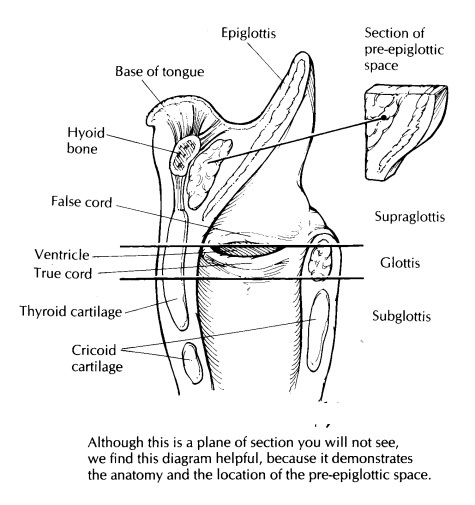
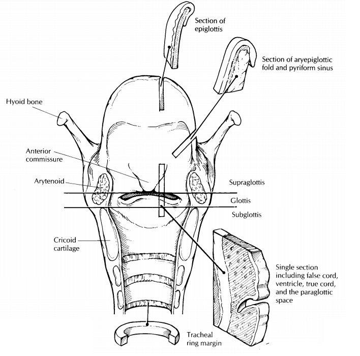
The most
important thing to remember about laryngal anatomy is that the larynx is
composed of three anatomic regions: the supraglottis, the glottis, and the
subglottis. The supraglottis is the portion of the larynx superior to the
ventri-cles. It is composed of the epiglottis, the aryte-noids, the
aryepiglottic folds, and the false cords. The glottis is composed of the true
vocal cords and their anterior and posterior attachments, the anterior and
posterior commissures. The sub-glottis begins 1 cm below the free edge of the
vocal cords and extends inferiorly to the trachea. These three anatomic regions
should be kept in mind throughout your description and dissec-tion of the
larynx.
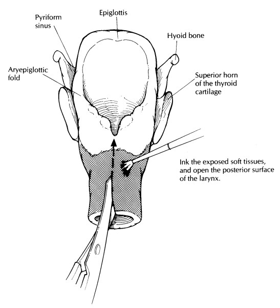
The
important mucosal landmarks to identify in the larynx are illustrated and
include the mucosa of the epiglottis, the aryepiglottic folds, the false vocal
cords, the ventricles, the true vocal cords, and the subglottis. Some specimens
may also include the base of the tongue with its over-lying mucosa. These
mucosal surfaces cover the cartilaginous framework of the larynx. This
framework includes the cartilage of the epiglot-tis, the thyroid cartilage, and
the cricoid cartilage. The epiglottis is attached to the thyroid carti-lage by
the thyroepiglottic ligament. The shield-shaped thyroid cartilage forms the
anterior and lateral walls of the larynx. The cricoid cartilage is shaped like
a signet ring, and it forms the posterior wall of the larynx. Situated in the
back of the larynx are the two arytenoid cartilages. These are pyramidal in
shape and rest along the upper border of the cricoid cartilage. Although the
hyoid bone is not technically part of the larynx, it is often included in
laryngectomy specimens.
Three
additional anatomic landmarks need to be defined because cancers that invade
these landmarks frequently escape from the larynx. The pre-epiglottic space is the triangular space ante-rior to the base
of the epiglottis. The pre-epiglottic space is filled with fatty connective
tissue, and it is bounded posteriorly by the epiglottis, inferi-orly by the
thyroepiglottic ligament, anteriorly by the thyrohyoid membrane, and superiorly
by the hyoepiglottic ligament. The paraglottic
space is a less well-defined area composed of loose connec-tive tissue,
which lies between the thyroid carti-lage and two membranes that form the
structural base for the vocal folds, the conus elasticus and quadrangular
membrane. The anterior commissure is
the anterior dense ligamentous attachment of the true vocal cords to the
thyroid cartilage. The thyroid cartilage lacks an internal perichon-drium;
therefore, carcinomas may invade the thyroid cartilage at the level of the
anterior com-missure. Carcinomas may also escape the larynx inferiorly via the
cricothyroid membrane.
Finally,
although the pyriform sinuses are tech-nically part of the hypopharynx, one
should be aware of them because they are frequently resected with the larynx.
The pyriform sinuses are small pouches that extend inferiorly from the
intersection of the aryepiglottic folds, glossoepig-lottic folds, and
pharyngeal wall. Depending on the size and location of the tumor, laryngeal
specimens may also include other portions of the hypopharynx (including the
posterior pharyn-geal wall and the pharyngo-esophageal junction) and the
thyroid gland.
Related Topics