Chapter: Medical Electronics : Radiological Equipments
Diagnostic X-Ray Equipments
DIAGNOSTIC
X-RAY EQUIPMENTS
X- rays are electromagnetic waves X-rays are
similar to light and sound waves. But the frequency and wavelength are
different.
·
Electromagnetic waves follow the relationship
v= fλ
Where
ν =
velocity
λ =
wavelength
f =
frequency
·
Electromagnetic wave is propagated in straight
line.
·
Electromagnetic wave follows the inverse square law
Intensity
α 1/ d2
Where d =
distance covered by the electromagnetic wave
·
Electromagnetic waves are not deflected by the
magnetic fields.
·
Electromagnetic waves produce interference
X Ray Radiations
X ray was
invented by Roentgen in 1895.
Two types
of X ray radiations are
·
Bremstrahlung radiation
·
Characteristic X ray radiation
X rays
are produced from the two different types of radiation that are listed above.
BREMSTRAHLUNG RADIATION
1. When the
fst moving electron enters into the orbit of anode material atom, its velocity
is continuously decreased due to scattering by the orbiting electrons. Thus the
loss of energy of that incident electrons will appear in the form of continuous
X rays or white rays.
2.
This type of radiation is used in medical
application based on the principle of energy absorption.
CHARACTERISTIC X RAY RADIATION
1.
It occurs when the incident electron ejects out of
the K shell or L shell e- in the anode material atom.Immediately the
higher orbit e- will falls into the vacancy to achieve equilibrium.
During its transition, the extra energy is emitted in the form of characteristic
X ray photon.
X ray tube:
·
X ray tube is similar to that of CRT except it
employs rotating anode. The speed of rotating anode varies from 3600 to 10000
rpm. Electrons are emitted from the cathode and focused on rotating anode and X
ray is produced. Only one percent of energy is converted into X ray and 99% is
converted into thermal or heat energy.
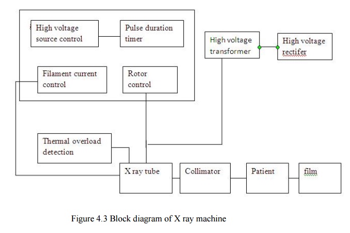
·
X ray machine has five different blocks
1. Power
supply
2. X ray
tube aluminium filter
3. Collimator
4. Bucky
diaphragm
5. Lead
shield
X ray
output can be obtained when electrons heat the anode
Q=
constant IB t VA2
IB
= Beam current
T =
exposure time
Q = X ray
output
VA
= Anode voltage in the order of 100 kV
Efficiency
η = X ray beam energy/ Electron beam energy
= 1.4 x
10-9 Z VA
Density
or darkness of the image is directly proportional to the amount of X rays that
penetrate the film
Contrast
is measure of the darkness of desired image.
Contrast
between two tissues = 10 log I1/I2 decibel
Power supply
·
X ray will have high voltage source and high
voltage transformer and high voltage rectifier. The power supply arrangement
will have the options for filament current control, rotor control, timer
control and thermal overload production circuit.
·
The various component in the X ray machine are used
to improve the the quality of the image, increase the contrast, improve the
resolution size and minimize the dose of X rays used on the patient.
·
The density or darkness of the image is
proportional to the amount of X rays that penetrate the film. Contrast is a
measure of darkness of the desired image, compare to its surroundings.
·
The good contrast in image will mainly depends on
the mass attenuation coefficient.
Aluminum filters:
The
emitted X rays wiil contain a broad range of frequency generally the aluminium
filters will observe the lower X ray frequency and hence the intensity of low
frequency X ray incident on the patient is reduced.
Collimator:
Collimator
is placed between the patient and aluminium filter. It is nothing but an
aperture .
diaphragm
which restricts the X ray beam falling on the patient. The necessary shape of X
ray beam is obtained only by collimator. A lamp and reflecting mirror
arrangement will makes a visible attern on the patient, so that the medical
attendant can tell where the X ray will strike. And this arrangement is used to
align or positioning the beam on the patient.
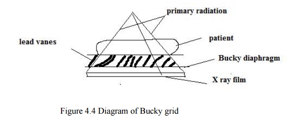
Bucky grid
·
It is used to reduce scattered radiation
·
The bucky grid is placed between the patient and
the film cassette to improve the sharpness of image.
Radiography
· X ray images developed by photography or photosensitive film
·
High resolution in images can be obtained
·
Wide range of contrast can be obtained
· Patient is not exposed to X rays during the examination of the X ray image
·
The patient dose is very low
· Permanent record is available
· The image can be obtained after developing the film and examination can be made before developing the film
·
Movement of organs cannot be observed
·
Efficient is more
Fluoroscopy
Fluoroscopy
is the method that provides real-time X ray imaging that is especially useful
for guiding a variety of diagnostic and interventional procedures.
The
ability of fluoroscopy to display motion is provided by a continuous series of
images produced at a maximum rate of 25-30 complete images per second. This is
similar to the way conventional television or video transmits images.
While the
X ray exposure needed to produce one fluoroscopic image is low (compared to
radiography), high exposures to patients can result from the large series of
images that are encountered in fluoroscopic procedures. Therefore, the total
fluoroscopic time is one of the major factors that determines the exposure to
the patient from fluoroscopy.

Because the X ray beam is usually moved over
different areas of the body during a procedure, there are two very different
aspects that must be considered. One is the area most exposed by the beam,
which results in the highest absorbed dose to that specific part of the skin
and to specific organs.The other is the total radiation energy imparted to the
patient’s body, which is related to the Kerma Area Product (KAP or PAK), a quantity that is easily
measurable.
The
absorbed dose to a specific part of the skin and other tissues is of concern in
fluoroscopy for two reasons: one is the need for minimizing the dose to
sensitive organs, such as the gonads and breast, by careful positioning of the
X ray beam and using shielding when appropriate.The second is the possible
incidence of the radiation beam to an area of the skin for a long time that can
result in radiation injuries in cases of very high exposure.
On the
other hand, the total radiation energy imparted to the patient’s body during a
procedure is closely related to the effective dose ) and to the risk of
radiation induced cancer.
In
fluoroscopy, as in all types of X ray imaging, the minimum exposure required to
form an image depends on the specific image information requirements.An
important characteristic of a fluoroscopic system is its sensitivity, i.e. the
amount of exposure required to produce images.
The use
of intensifier tubes and more modern digital flat panel receptors make it
possible to optimize the balance of patient exposure with image quality so as
not to expose the patient to unnecessary radiation.Non-intensified fluoroscopy
with just a fluorescent screen for a receptor should not be used because of the
excessive exposure to the patient.
Applications of X ray
·
X ray is used to visualize skeletal structure
·
X ray is used to take chest radiograph
·
Bronchography
·
Heart examinations are performed by taking frontal
and lateral X ray film images
·
Gastro intestinal tract can be imaged by using X
ray
·
Urinary tract can be examined by using X rays
Angiography
· Angiography is a special X ray imaging technique through which high contrasts can be obtained.
·
The outline of the blood vessels are visible in
angiogram
· Angiocardiography: It mean study of heart
·
Cerebral
angiography: It mean study of brain
·
Bronchography: It maen
study of lungs
·
Nephro
angiography: it mean study of kidney
Related Topics