Zoology - Connective Tissue | 11th Zoology : Chapter 3 : Tissue Level of Organisation
Chapter: 11th Zoology : Chapter 3 : Tissue Level of Organisation
Connective Tissue
Connective
Tissue
Connective
tissue develops from the mesoderm and is widely distributed in the body. There
are four main classes of connective tissues.They are connective tissue (which
includes fat and the fibrous tissue of ligaments), cartilage, bones and blood.
Major functions of connective tissues are binding and support, protection,
insulation and transportation of substances.
Components of connective tissue
All
connective tissues consist of three main componentsnamelyfibres,
groundsubstance and cells. The ‘Fibres’ of connective tissue provide support.
Three types of fibres are found in the connective tissue matrix. They are
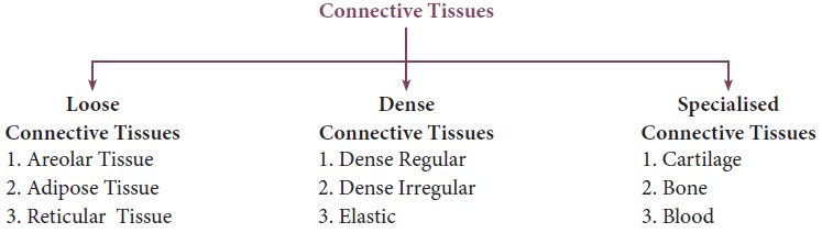
collagen, elastic and reticular fibres. Connective tissue are of two types
namely, Loose connective tissues (Areolar, Adipose and Reticular) and Dense
connective tissues (dense regular, dense irregular and elastic). Specialized
connective tissues include cartilage, bone and blood.
![]()
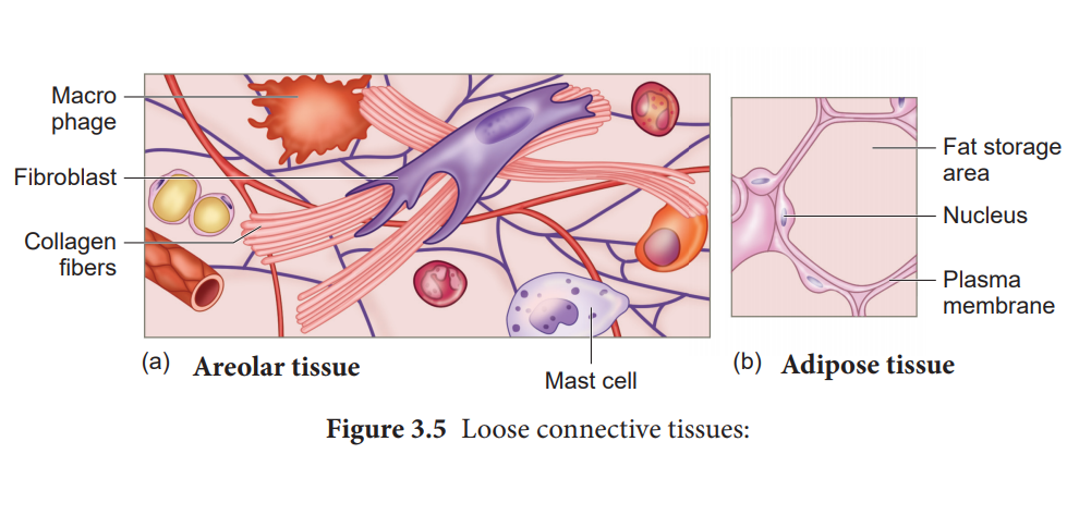
Loose connective tissues
In this
tissue the cells and fibres are loosely arranged in a semi fluid ground
substances. For example the Areolar connective tissue beneath the skin acts as
a support framework for epithelium and acts as a reservoir of water and salts
for the surrounding body tissues, hence aptly called tissue fluid. It contains
fibroblasts, macrophages, and mast cells (Figure 3.5).
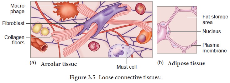
Adipose
tissue is similar to areolar tissue in structure and function and located
beneath theskin. Adipocytes commonly called adipose or fat cells predominate
and account for 90% of this tissue mass. The cells of this tissue store fats
and the excess nutrients which are not utilised immediately are converted to
fats and are stored in tissues. Adipose tissue is richly vascularised
indicating its high metabolic activity. While fasting, these cells maintain life
by producing and supplying energy as fuel. Adipose tissues arealso found in
subcutaneous tissue, surrounding the kidneys, eyeball, heart,
etc. Adipose tissue is called ‘white fat’ or white adipose tissue. The adipose
tissue which contains abundant mitochondria is called ‘Brown fat’ or Brown
adipose tissue. White fat stores nutrients whereas brown fat is used to heat
the blood stream to warm the body. Brown fat produces heat by non-shivering
thermogenesis in neonates.
Reticular
connective tissue resembles areolar connective tissue, but, the matrix is
filled with fibroblasts called reticular cells. It forms an internal framework
(stroma) that supports the blood cells (largely lymphocytes) in the lymph
nodes, spleen and bone marrow.
Dense connective tissues (connective tissue proper)
Fibres
and fibroblasts are compactly packed in the dense connective tissues.
Orientation of fibres show a regular or irregular pattern and is called dense
regular and dense irregular tissues. Dense
regular connective tissues primarily contain collagen fibres in rows
between many parallel bundles of tissues and a few elastic fibres. The major
cell type is fibroblast. It attaches
muscles and bones and withstands great tensile stress when pulling force is
applied in one direction. This connective tissue is present in tendons, that attach skeletal muscles
to bones and ligaments attach one bone to another. Dense irregular connective tissues have bundles of thick collagen
fibres and fibroblasts which are arranged irregularly. The major cell type is
the fibroblast. It is able to
withstand tension exerted in many directions and provides structural strength.
Some elastic fibres are also present. It is found in the skin as the leathery
dermis and forms fibrous capsules of organs such as kidneys, bones, cartilages,
muscles, nerves and joints. Elastic
connectivetissue contains high proportion of elastic fibres. It allows
recoil of tissues following stretching. It maintains the pulsatile flow of
blood through the arteries and the passive recoil of lungs following
inspiration. It is found in the walls of large arteries; ligaments associated
with vertebral column and within the walls of the bronchial tubes.
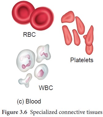
Specialised connective tissues are
classified as cartilage, bones and blood. The intercellular material of cartilage
is solidand pliable and resists compression. Cells of this tissue
(chondrocytes) are enclosed in small cavities within the matrix secreted by
them (Figure 3.6). Most of the cartilages in vertebrate embryos are replaced by
bones in adults. Cartilage is present in the tip of nose, outer ear joints, ear
pinna, between adjacent bones of the vertebral column, limbs and hands in
adults.
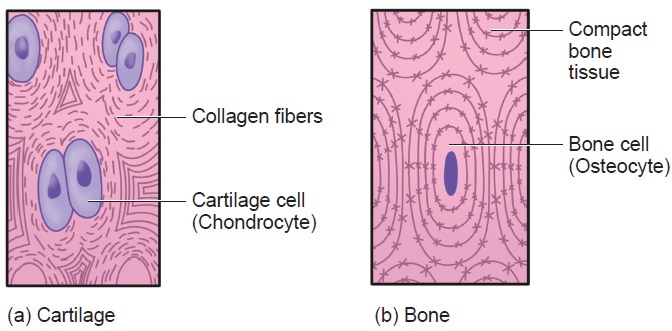
Bones have a hard and non-pliable ground substance rich in calcium
salts
It is the main tissue that
provides structural frame to the body. Bones support and protect softer tissues
and organs. The bone cells (osteocytes) are present in the spaces called lacunae.
Limb bones, such as the long bones of the legs, serve weight-bearing functions.
They also interact with skeletal muscles attached to them to bring about
movements. The bone marrow in some bones is the site of production of blood
cells.
Blood is the fluid connective tissue
containing plasma, red blood cells (RBC), white blood cells (WBC) and
platelets. It functions as the transport medium for the cardiovascular system,
carrying nutrients, wastes, respiratory gases throughout the body.
Related Topics