Chapter: Human Nervous System and Sensory Organs : Brain's Cerebrovascular and Ventricular Systems
Veins - Cerebrovascular and Ventricular Systems

Veins
The major veins lie on the surface of the brain in the subarachnoid space; some deep veins run beneath the ependyma. The cerebral veins do not possess valves. They ex-hibit considerable variations with respect to course and drainage. Quite often there are several small vessels instead of a single major vein. The cerebral veins are divided into two groups: the superficial cerebral veins which drain the blood into the sinuses of the dura mater , and the deep cerebralveins which drain the blood into thegreatcerebral vein (great vein of Galen).
Superficial Cerebral Veins
We distinguish between the group of supe-rior cerebral veins and the group of inferior cerebral veins. The superior cerebral veins (AC1), totaling about 10 – 15 veins, collect the blood from the frontal and parietal lobes and carry it into the superior sagittal sinus (BC2). They run within the subarachnoid space and empty into the lateral lacunae (BC3), pouchlike cavities of the superior sagittal sinus. For a short distance, they pass through the subdural space. Here, the thin-walled veins can easily rupture during head injury and bleed into the subdural space (subdural hematoma). Strangely, the veins empty into the superior sagittal sinus at an oblique angle against the dominating blood flow in the sinus.
The inferior cerebral veins receive the blood from the temporal lobe and from the basal regions of the occipital lobe; they empty into the transverse sinus and the su-perior petrosal sinus. The largest and mostconsistent of these veins is the superficialmiddle cerebral vein (AC4) located in thelateral sulcus; it often consists of several venous trunks. It drains the blood from most of the lateral aspect of the hemisphere into the cavernous sinus.
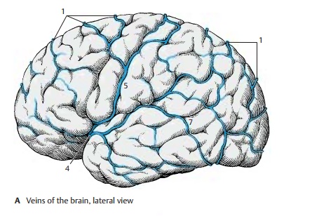
The superior and inferior cerebral veins are interconnected only by a few anastomoses. The most important one is the superior anastomotic vein (Trolard’s vein) (AC5); itempties into the superior sagittal sinus and is connected with the superficial middle cerebral vein. The central vein (C6) located in the central sulcus may also form anasto-moses with the middle cerebral vein. The inferior anastomotic vein (Labbé’s vein,Browning ’s vein) (AC7) connects the super-ficial middle cerebral vein with the trans-verse sinus.
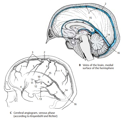
Carotid angiogram (venous phase).A dia-gram of the venous phase of a carotid angio-gram is shown in C (for arterial phase, see p. 272). A radiograph taken only seconds after injection of the contrast medium shows its drainage via the venous vascular tree. Superficial and deep veins are seen in a single plane.
C9 Internal cerebral vein.
C10 Thalamostriate vein (terminal vein).
C11 Vein of the septum pellucidum
C12 Interventricular foramen (foramen of Monro).
C13 Basal vein (Rosenthal’s vein).
BC14 Straight sinus.
BC15 Inferior sagittal sinus.
BC16 Confluence of sinuses.
Deep Cerebral Veins (A, B)
The deep cerebral veins collect the blood from the diencephalon, the deep structures of the hemispheres, and the deep white matter. In addition, there are thin transcerebral veins running along the fibers of the corona radi-ata from the outer white matter and from the cortex. They connect the superficial drainage areas with the deep ones. The deep cranial veins empty their blood into the great cerebral vein (great vein of Galen). Thedrainage system of the deep veins is there-fore also known as the system of the great cerebral vein.
The great cerebral vein (AB1) is a short vascular trunk formed by the confluence of four veins, namely, the two internal cerebralveins and the two basal veins. It curvesaround the splenium of the corpus callosum and empties into the straight sinus. Veins from the surface of the cerebellum and from the occipital lobe (B2) may drain into it.
The basal vein (Rosenthal’s vein) (AB3) arises at the anterior perforated substance (A4) by junction of the anterior cerebral vein and the deep middle cerebral vein.
The anterior cerebral vein (A5) receives its blood from the anterior two-thirds of the corpus callosum and the adjacent convolu-tions. It extends around the genu of the cor-pus callosum to the base of the frontal lobe. The deep middle cerebral vein (A6) arises in the insular area and receives veins from the basal parts of putamen and globus pallidus.
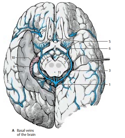
The basal vein crosses the optic tract and as-cends in the cisterna ambiens around the cerebral peduncle (A7) to below the splenium, where it empties into the great cerebral vein. Along its course it receives numerous venous tributaries, namely, veins from the optic chiasm and the hypo-thalamus, the interpeduncular vein (A8), the inferior choroid vein (A9) from the choroidplexus (A10) of the inferior horn, and veins from the internal segment of the globus pal-lidus and from the basal parts of the thalamus.
The internal cerebral vein (AB11) arises at the interventricular foramen (foramen of Monro) by junction of the vein of the septumpellucidum, the thalamostriate vein, and the superior choroid vein.
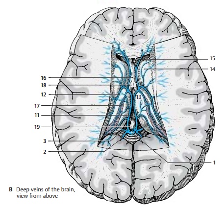
The thalamostriate vein (terminal vein) (B12) runs in the terminal sulcus between thalamus (B13) and caudate nucleus (B14) in rostral direction to the interventricular foramen. It receives venous tributaries from the caudate nucleus, from the adjacent white matter, and from the lateral corner of the lateral ventricle. The vein of the septumpellucidum (B15) receives venous branchesfrom the septum pellucidum (B16) and from the deep frontal white matter. The choroidvein (B17) runs with the choroid plexus tothe inferior horn. In addition to the vessels of the plexus, it receives the veins from the hippocampus and from the deep temporal white matter.
The internal cerebral vein extends from the interventricular foramen across the medial surface of the thalamus at the margin of the roof of the diencephalon to the region of the pineal gland, where it unites with the con-tralateral internal cerebral vein and the basal veins to form the great cerebral vein. Along its course it receives tributaries from the fornix (B18), from the dorsal parts of the thalamus, from the pineal gland (epiphysis) (B19) and, variably, from the deep white matter of the occipital lobe.In summary, the dorsal parts of thalamus, pallidum, and striatum drain into the inter-nal cerebral vein, while the ventral parts drain into the basal vein.
Clinical Note: Obstruction of a cerebral veincauses congestion and hemorrhage in the affected region. In case of birth trauma, rupture of the thalamostriate vein in newborns may lead to hemorrhage into the ventricles.
A20 Superficial middle cerebral vein.
Related Topics