Chapter: Pathology: Fundamentals of Pathology
Overview of Pathology
OVERVIEW OF PATHOLOGY
Definitions
The study
of the essential nature of disease, including symptoms/signs, patho-genesis,
complications, and morphologic consequences including structural and functional
alterations in cells, tissues, and organs
The study
of all aspects of the disease process focusing on the pathogenesis leading to classical
structural changes (gross and histopathology) and molecu-lar alterations
The
etiology (cause) of a disease may be
genetic or environmental. The pathogen-esis
of a disease defines the temporal sequence and the patterns of cellular
injurythat lead to disease. Morphologic
changes of the disease process include both gross changes and microscopic
changes. The clinical significance
of a disease relates to its signs and symptoms, disease course including
complications, and prognosis.
Methods Used
Gross examination of
organs on exam questions has 2 major components: identify-ing the organ and
identifying the pathology. Useful gross features include consid-eration of
size, shape, consistency, and color.
Microscopic examination of tissue
In
light microscopic examination of tissue, hematoxylin
and eosin (H&E) is considered the gold standard stain and is used
routinely in the initial micro-scopic examination of pathologic specimens.
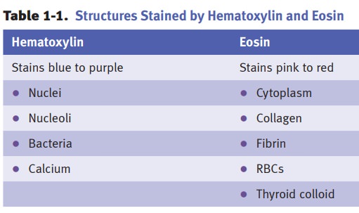
The
common denominator of the features shown in Table 1-1 is that hematoxylin binds
nucleic acids and calcium salts, while eosin stains the majority of proteins
(both extracellular and intracellular).
·
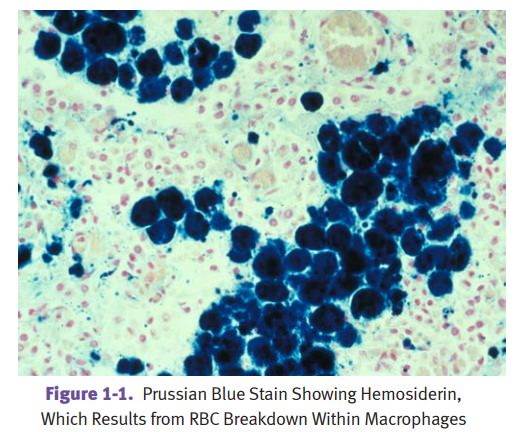
Other
histochemical stains (chemical reactions): Prussian blue
(stains iron),Congo red (stains amyloid), acid fast (Ziehl-Neelsen, Fite)
(stains acid-fast bacilli), periodic acid-Schiff (PAS, stains high carbohydrate
content mol-ecules), Gram stain (stains bacteria), trichrome (stains cells and
connective tissue), and reticulin (stains collagen type III molecules).
·
Immunohistochemical
(antibody) stains include cytokeratin (stains
epithe-lial cells), vimentin (stains cells of mesenchymal origin except the 3
muscle types; stains many sarcomas), desmin (stains smooth, cardiac, and
skeletal myosin), prostate specific antigen, and many others.
Ancillary techniques include immunofluorescence microscopy (IFM),
typicallyused for renal and autoimmune disease, and transmission electron microscopy(EM), used for renal disease,
neoplasms, infections, and genetic disorders.
Molecular techniques include
protein electrophoresis, Southern and Western blots,polymerase chain reaction
(PCR), and cytogenetic analysis (karyotyping, in situ hybridization studies).
Summary
Pathology
is the study of disease and concerns itself with the etiology, pathogenesis,
morphologic changes, and clinical significance of different diseases.
Gross
examination of organs involves identifying pathologic lesions by evaluating
abnormalities of size, shape, consistency, and color.
Tissue
sections stained with hematoxylin (nucleic acids and calcium salts) and eosin
(most proteins) are used for routine light microscopic examination.
Additional
techniques that pathologists use to clarify diagnoses include histochemical
stains, immunohistochemical stains, immunofluorescence microscopy, transmission
electron microscopy, and molecular techniques.
Related Topics