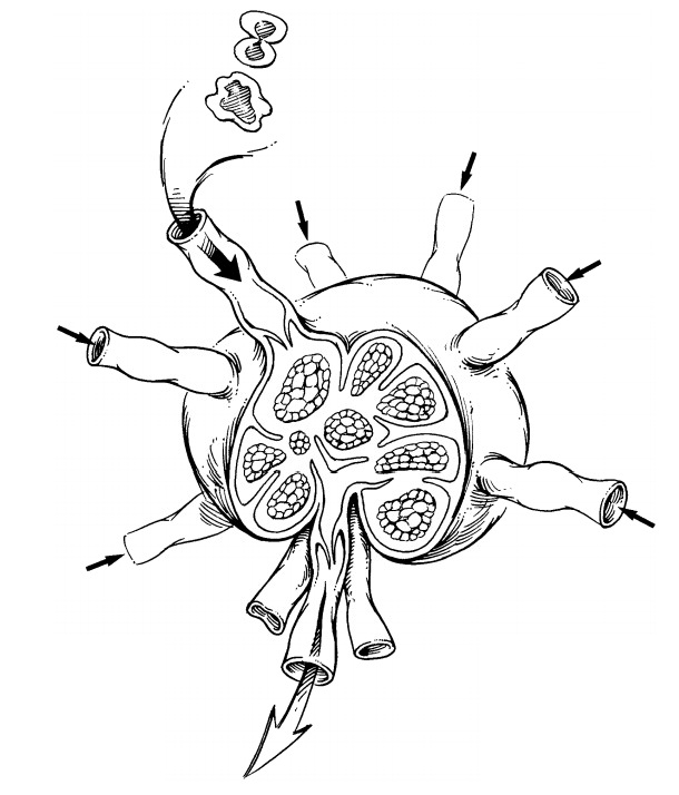
Chapter: Surgical Pathology Dissection : Lymph Nodes
Lymph Node Dissections
Lymph Nodes
Metastatic
spread to lymph nodes carries pro-found treatment and prognostic implications
for patients with carcinomas, melanomas, or any other malignant neoplasm with
metastatic po-tential. Accordingly, assessment of lymph node specimens is every
bit as important as evaluating a neoplasm at its primary site.

Lymph Node Dissections
A
general approach to sampling lymph nodes is described earily, and the
organ-specific approach to the dissection of regional lymph nodes is detailed
in each organ-specific.
Lymph
nodes are easiest to appreciate in the fresh state before subtle distinctions
in tissue den-sity are obscured by tissue fixation. Submersion of specimens in
certain clearing agents may be help-ful in eliminating the bulk of adipose
tissue, but this is an unnecessary and time-consuming step that does not
improve on meticulous exam-ination of the fresh tissues. When appropriate, the
anatomic levels of the lymph nodes should be maintained and separately reported
in the sur-gical pathology report (e.g., colectomy speci-mens and neck
dissection specimens). In cases where important anatomic landmarks do not
accompany the specimen, it may not be possible to identify levels without the
help of the sub-mitting surgeon.
Although
tedious, the objective of any lymph node dissection for metastatic disease is
nothing less than detection and processing of every lymph node contained in the
specimen. There is practically no role for representative nodal sam-pling when
searching for metastatic spread. Each lymph node identified should be submitted
for microscopic examination. If tumor is grossly vis-ible, a section that
includes the tumor suffices. If tumor is not grossly visible, the lymph node
should be sectioned in 3- to 4-mm slices and all of the sections submitted for
microscopic eval-uation. You can slice the lymph node along its long or short
axis, but longitudinal sections are generally preferable, as this minimizes the
number of slices. No single cassette should contain slices from more than one
lymph node unless colored inks are used to distinguish different lymph nodes.
For these nonsentinel lymph node dissections, one hematoxylin and eosin
(H&E)-stained section per tissue block is adequate.
Related Topics