Chapter: 10th Science : Chapter 16 : Plant and Animal Hormones
Human Endocrine Glands
Human Endocrine Glands
Endocrine glands in
animals possess a versatile communication system to coordinate biological
functions. Exocrine glands and endocrine glands are two kinds of glands found
in animals. Endocrine glands are found in different regions of the body of
animals as well as human beings. These glands are called ductless glands.
Their secretions are called hormones which are produced in minute
quantities. heT secretions diffuse into the blood stream and are carried to
the distant parts of the body. They act on specific organs which are referred
as target organs.
Exocrine glands have
specific ducts to carry their secretions e.g. salivary glands, mammary glands,
sweat glands.
Endocrine glands present
in human and other vertebrates are
a)
Pituitary gland
b)
Thyroid gland
c)
Parathyroid gland
d)
Pancreas (Islets of Langerhans)
e)
Adrenal gland (Adrenal cortex and Adrenal medulla)
f)
Gonads(Testes and Ovary)
g)
Thymus gland
1. Pituitary Gland
![]() The pituitary gland or hypophysis is
a pea shaped compact mass of cells located at the base of the midbrain attached
to the hypothalamus by a pituitary stalk. The pituitary
The pituitary gland or hypophysis is
a pea shaped compact mass of cells located at the base of the midbrain attached
to the hypothalamus by a pituitary stalk. The pituitary
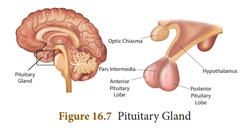
They are the anterior
lobe (adenohypophysis) and the posterior lobe (neurohypophysis).
The intermediate lobe is non-existent in humans.
The pituitary gland
forms the major endocrine gland in most vertebrates. It regulates and controls
other endocrine glands and so is called as the “Master gland”.
Hormones secreted by the anterior lobe (Adenohypophysis) of pituitary
The anterior pituitary
is composed of different types of cells and secrete hormones which stimulates the
production of hormones by other endocrine glands. The hormones secreted by
anterior pituitary are
a)
Growth Hormone
b)
Thyroid stimulating Hormone
c)
Adrenocorticotropic Hormone
d)
Gonadotropic Hormone which comprises the Follicle Stimulating
Hormone and Luteinizing Hormone
e)
Prolactin
a. Growth hormone (GH)
GH promotes the
development and enlargement of all tissues of the body. It stimulates the
growth of muscles, cartilage and long bones. It controls the cell metabolism.
The improper secretion
of this hormone leads to the following conditions.
Dwarfism: It is caused by
decreased secretion of growth hormone in children. The characteristic
features are stunted growth, delayed skeletal formation and mental disability.
Gigantism: Oversecretion of growth
hormone leads to gigantism in children. It is characterised by overgrowth
of all body tissues and organs. Individuals attain abnormal increase in height.
Acromegaly: Excess secretion of
growth hormone in adults may lead to abnormal enlargement of head, face,
hands and feet.
b. Thyroid stimulating hormone (TSH)
TSH controls the growth
of thyroid gland, coordinates its activities and hormone secretion.
c. Adrenocorticotropic hormone (ACTH)
ACTH stimulates adrenal
cortex of the adrenal gland for the production of its hormones. It also
influences protein synthesis in the adrenal cortex.
d. Gonadotropic hormones (GTH)
The gonadotropic
hormones are follicle stimulating hormone and luteinizing hormone which are
essential for the normal development of gonads.
Follicle stimulating hormone
(FSH)
In male, it stimulates
the germinal epithelium of testes for formation of sperms. In female it
initiates the growth of ovarian follicles and its development in ovary.
Luteinizing hormone (LH)
In male, it promotes the
Leydig cells of the testes to secrete male sex hormone testosterone. In female,
it causes ovulation (rupture of mature graafian follicle), responsible for the
development of corpus luteum and production of female sex hormones estrogen and
progesterone.
e. Prolactin (PRL)
PRL is also called lactogenic
hormone. This hormone initiates development of mammary glands during
pregnancy and stimulates the production of milk after child birth.
Hormones
secreted by the posterior lobe (Neurohypophysis) of pituitary
The hormones secreted by
the posterior pituitary are
1.
Vasopressin or Antidiuretic hormone
2.
Oxytocin
a. Vasopressin or Antidiuretic hormone (ADH)
In kidney tubules it
increases reabsorption of water. It reduces loss of water through urine and
hence the name antidiuretic hormone.
Deficiency of ADH
reduces reabsorption of water and causes an increase in urine output
(polyuria). This deficiency disorder is called Diabetes insipidus.
b. Oxytocin
It helps in the
contraction of the smooth muscles of uterus at the time of child birth and milk
ejection from the mammary gland after child birth.
2. Thyroid Gland
The thyroid gland is
composed of two distinct lobes lying one on either side of the trachea. The two
lobes are connected by means of a narrow band of tissue known as the isthmus.
This gland is composed of glandular follicles and
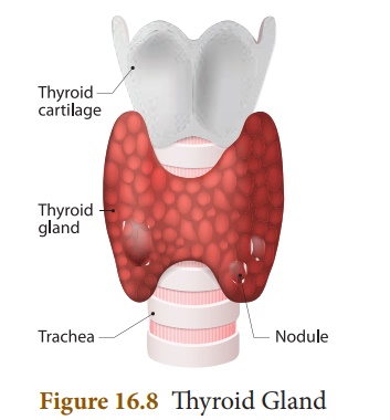
lined by cuboidal
epithelium.The follicles are fsilled with colloid material called thyroglobulin.
An amino acid tyrosine
and iodine are involved in the formation of thyroid hormone. The
hormones secreted by the thyroid gland are
a. Triiodothyronine (T3)
b. Tetraiodothyronine or Thyroxine (T4)
Functions of thyroid hormones
The functions of thyroid
hormones are
·
Production of energy by maintaining the Basal Metabolic Rate (BMR) of the body.
·
Helps to maintain normal body temperature.
·
Influences the activity of central nervous system.
·
Controls growth of the body and bone formation.
·
Essential for normal physical, mental and personality development
.
·
It is also known as personality hormone.
·
Regulates cell metabolism.
Thyroid Dysfunction
When the thyroid gland
fails to secrete the normal level of hormones, the condition is called thyroid
dysfunction. It leads to the following conditions
Hypothyroidism
It is caused due to the
decreased secretion of the thyroid hormones. The abnormal conditions are simple
goitre, cretinism and myxoedema.
Goitre
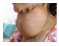
It is caused due to the
inadequate supply of iodine in our diet. This is commonly prevalent in
Himalayan regions due to low level of iodine content in the soil. It leads to
the enlargement of thyroid gland which protrudes as a marked swelling in the
neck and is called as goitre.
Cretinism
It is caused due to
decreased secretion of the thyroid hormones in children. The conditions are
stunted growth, mental defect, lack of skeletal development and deformed bones.
They are called as cretins.
Myxoedema
It is caused by
deficiency of thyroid hormones in adults. They are mentally sluggish, increase
in body weight, puffiness of the face and hand, oedematous appearance.
Hyperthyroidism
It is caused due to the
excess secretion of the thyroid hormones which leads to Grave’s disease. The
symptoms are protrusion of the eyeballs (Exopthalmia), increased metabolic
rate, high body temperature, profuse sweating, loss of body weight and nervousness.
3. Parathyroid Gland
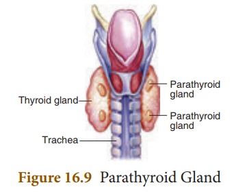
The parathyroid glands
are four small oval bodies that are situated on the posterior surface of the
thyroid lobes. The chief cells of the gland are mainly concerned with
secretion of parathormone.
Functions of Parathormone
The parathormone
regulates calcium and phosphorus metabolism in the body.They act on bone,
kidney and intestine to maintain blood calcium levels.
Parthyroid Dysfunction
The secretion of
parathyroid hormone can be altered due to the following conditions.
Removal of parathyroid
glands during thyroidectomy (removal of thyroid) causes decreased secretion of
parathormone. The conditions are
·
Muscle spasm known as Tetany (sustained contraction of
muscles in face, larynx, hands and feet).
·
Painful cramps of the limb muscles.
4. Pancreas (Islets of Langerhans)
Pancreas is an
elongated, yellowish gland situated in the loop of stomach and duodenum. It is exocrine
and endocrine in nature. The exocrine pancreas secretes pancreatic juice
which plays a role in digestion while, the endocrine portion is made up of
Islets of Langerhans.
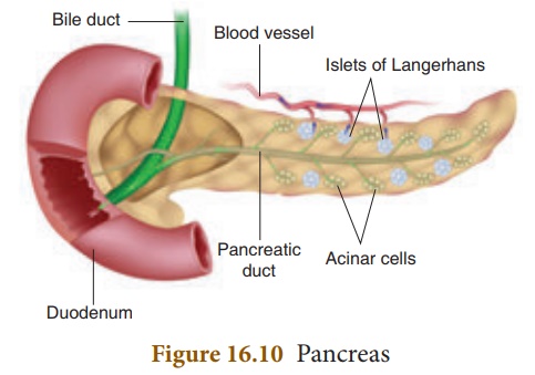
The Islets of Langerhans
consists of two types of cells namely alpha cells and beta cells. The alpha
cells secrete glucagon and beta cells secrete insulin.
Functions of Pancreatic hormones
A balance between
insulin and glucagon production is necessary to maintain blood glucose
concentration.
Insulin
·
Insulin helps in the conversion of glucose into glycogen which is
stored in liver and skeletal muscles.
·
It promotes the transport of glucose into the cells.
·
It decreases the concentration of glucose in blood.
Glucagon
·
Glucagon helps in the breakdown of glycogen to glucose in the
liver.
·
It increases blood glucose levels.
Diabetes mellitus
The deficiency of
insulin causes Diabetes mellitus. It is characterised by
·
Increase in blood sugar level (Hyperglycemia).
·
Excretion of excess glucose in the urine (Glycosuria).
·
Frequent urination (Polyuria).
·
Increased thirst (Polydipsia).
·
Increase in appetite (Polyphagia).
5. Adrenal Gland
The adrenal glands are
located above each kidney. They are also called supra renal glands.
The outer part is the
adrenal cortex and the inner part is the adrenal medulla. The two distinct
parts are structurally and functionally different.
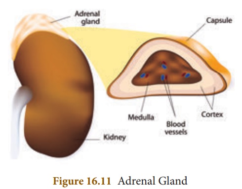
Adrenal Cortex
The adrenal cortex
consists of three layers of cells. They are zona glomerulosa, zona
fasciculata and zona reticularis
Hormones of Adrenal Cortex
The hormones secreted by
the adrenal cortex are corticosteroids. They are classified into
a. Glucocorticoids
b. Mineralocorticoids
Functions of adrenocortical hormones
Glucocorticoids
The glucocorticoids
secreted by the zona fasciculata are cortisol and corticosterone
·
They regulate cell metabolism.
·
It stimulates the formation of glucose from glycogen in the liver.
·
It is an anti-inflammatory and anti-allergic agent.
Mineralocorticoids
The mineralocorticoids
secreted by zona glomerulosa is aldosterone
·
It helps to reabsorb sodium ions from the renal tubules.
·
It causes increased excretion of potassium ions.
·
It regulates electrolyte balance, body fluid volume, osmotic
pressure and blood pressure.
Adrenal Medulla
The adrenal medulla is
composed of chromaffin cells .They are richly supplied with sympathetic
and parasympathetic nerves.
Hormones of Adrenal Medulla
It secretes two hormones
namely
a. Epinephrine (Adrenaline)
b. Norepinephrine (Noradrenaline)
They are together called
as “Emergency hormones”. It is produced during conditions of
stress and emotion. Hence it is also referred as “flight, fright and fight
hormone”.
Functions of adrenal medullary hormones
Epinephrine (Adrenaline)
·
It promotes the conversion of glycogen to glucose in liver and
muscles.
·
It increases heart beat and blood pressure.
·
It increases the rate of respiration by dilation of bronchi and
trachea.
·
It causes dilation of the pupil in eye.
·
It decreases blood flow through the skin.
Norepinephrine
(Noradrenalin)
Most of its actions are similar to those of epinephrine.
6. Reproductive Glands (Gonads)
The sex glands are of
two types the testes and the ovaries. The testes are present in
male, while the ovaries are present in female.
Testes
Testes are the
reproductive glands of the males. They are composed of seminiferous tubules,
Leydig cells and Sertoli cells. Leydig cells form the endocrine
part of the testes. They secrete the male sex hormone called testosterone.
Functions of testosterone
·
It influences the process of spermatogenesis.
·
It stimulates protein synthesis and controls muscular growth.
·
It is responsible for the development of secondary sexual
characters (distribution of hair on body and face, deep voice pattern, etc).
Ovary
The ovaries are the
female gonads located in the pelvic cavity of the abdomen. They secrete the
female sex hormones
·
Estrogen
·
Progesterone
Estrogen is produced by the
Graafian follicles of the ovary and progesterone from the corpus
luteum that is formed in the ovary from the ruptured follicle during
ovulation.
Functions of estrogens
·
It brings about the changes that occur during puberty.
·
It initiates the process of oogenesis.
·
It stimulates the maturation of ovarian follicles in the ovary.
·
It promotes the development of secondary sexual characters (breast
development, high pitched voice etc).
Functions of progesterone
·
It is responsible for the premenstrual changes of the uterus.
·
It prepares the uterus for the implantation of the embryo.
·
It maintains pregnancy.
·
It is essential for the formation of placenta.
![]()
![]()
7. Thymus Gland
Thymus is partly an
endocrine gland and partly a lymphoid gland. It is located in the upper part of
the chest covering the lower end of trachea. Thymosin is the hormone
secreted by thymus.
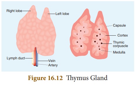
Functions of Thymosin
·
It has a stimulatory effect on the immune function.
·
It stimulates the production and differentiation of lymphocytes.
Related Topics