Chapter: Pathology: Inflammation
Chronic Inflammation
CHRONIC INFLAMMATION
Causes
of chronic inflammation include the following:
·
Following a bout of acute
inflammation
·
Persistent infections
·
Infections with certain organisms,
including viral infections, mycobacteria, parasitic infections, and fungal
infections
·
Autoimmune diseases
·
Response to foreign material
·
Response to malignant tumors

There
are several important cells in chronic
inflammation.
·
Macrophages
are derived from blood monocytes. Tissue-based
macrophages(life span in connective tissue compartment is 60–120 days) are
found in connec-tive tissue (histiocyte), lung (pulmonary alveolar
macrophages), liver (Kupffer cells), bone (osteoclasts), and brain (microglia).
During inflammation circu-lating monocytes emigrate from the blood to the
periphery and differentiate into macrophages.
Respond
to chemotactic factors: C5a, MCP-1, MIP-1α,
PDGF, TGF-β
Secrete
a wide variety of active products (monokines)
May
be modified into epithelioid cells in granulomatous processes
·
Lymphocytes
include B cells and plasma cells, as well as T cells.
Lymphotaxinis the lymphocyte chemokine.
·
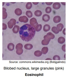
Eosinophils
play an important role in parasitic infections and
IgE-mediatedallergic reactions. The eosinophilic chemokine is eotaxin.
Eosinophil granules contain major basic protein, which is toxic to parasites.
·
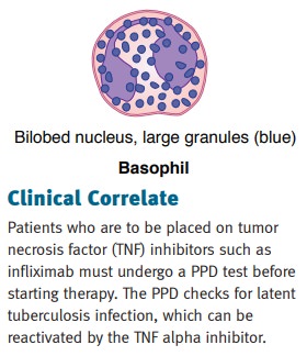
Basophils
contain similar chemical mediators as mast cells in their
granules.Mast cells are present in high numbers in the lung and skin. Both
basophils and mast cells play an important role in IgE-mediated reactions
(allergies and anaphylaxis) and can release histamine.
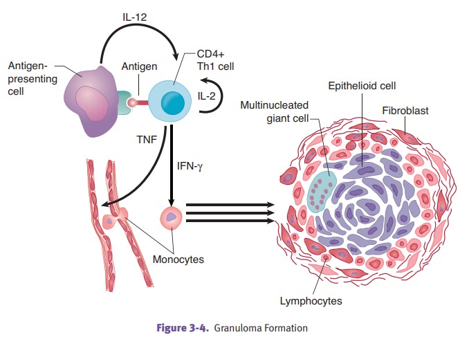
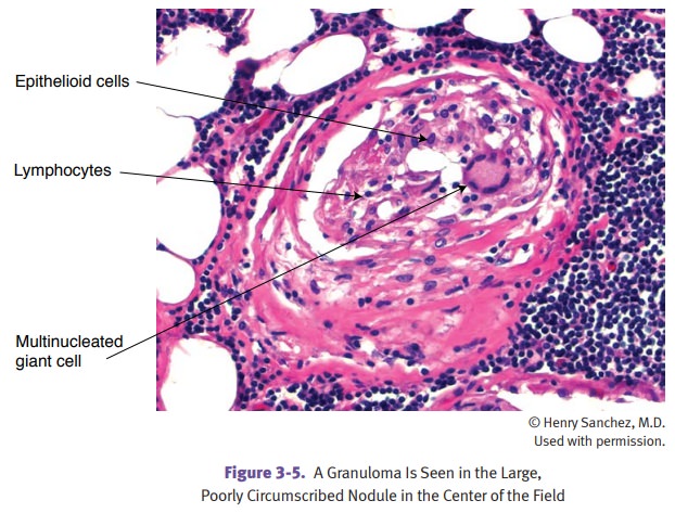
Chronic granulomatous inflammation is
a specialized form of chronic inflamma-tion characterized by small aggregates
of modified macrophages (epithelioid cells and multinucleated giant cells)
usually populated by CD4+ Th1 lymphocytes.
Composition
of a granuloma is as follows:
·
Epithelioid cells, located
centrally, form when IFN-γ
transforms macrophages to epithelioid cells. They are enlarged cells with
abundant pink cytoplasm.
·
Multinucleated giant cells, located
centrally, are formed by the fusion of epi-thelioid cells. Types include
Langhans-type giant cell (peripheral arrangement of nuclei) and foreign body
type giant cell (haphazard arrangement of nuclei).
·
Lymphocytes and plasma cells are
present at the periphery.
·
Central necrosis occurs in
granulomata due to excessive enzymatic breakdown and is commonly seen in Mycobacterium tuberculosis infection as
well as fun-gal infections and a few bacterial infections. Because of the
public health risk of tuberculosis, necrotizing granulomas should be considered
tuberculosis until proven otherwise.

Granulomatous diseases include
tuberculosis (caseating granulomas), cat-scratchfever, syphilis, leprosy,
fungal infections (e.g., coccidioidomycosis), parasitic infec-tions (e.g.,
schistosomiasis), foreign bodies, beryllium, and sarcoidosis.

