Chapter: Biology: Animal Tissue, Organ and Organ System
Types of Tissues
Types of Tissues :
On the basis of number of cells, characteristic and the presence or absence of the intercellular materials or matrix secreted by cells, the tissue is mainly divided into four categories.
1. The Epitherlial Tissue.
2. The Connective Tissue.
3. The Muscular Tissue.
4. The Nerve Tissue.
1. Epithelial Tissue
Structural Characteristics, Function and Location of Epithelial Tissue:
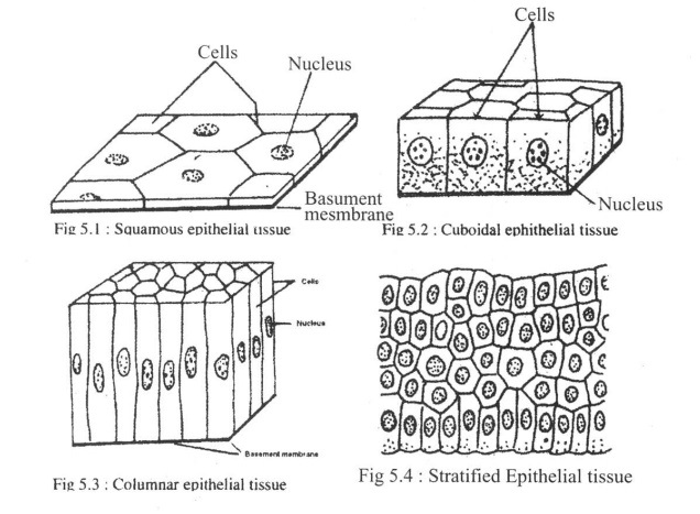
Structural Characterisics : The cells of the epithelial tissue lie closely or side byside on a basement membrane. There is no matrix in this tissue. On the basis of cell size, location in the animal body and nature of work, this tissue is of three types, sch as;
a. Squamous Epithelial Tissue: Cells of this tissue are flat like scales; rnucleusis large (Fig- 5.1)
Example: Wall of the Bowman's capsule of Kindey.
Function: Mainly filtration and covering.
b. Cuboidal Epithelial Tissue: Cells of this tissue are cuboidal, as the length,breadth and height of the cells are nearly equal (Fig-5.2).
Example: Collecting tubules of the kidney. Function: Mainly absorption and covering.
c. Columnar Epithelial Tissue : Cells of this tissue are narrow and elongated like acolumn (Fig-5.3)
Example: On the internal wall of intestine.
Function: Mainly secretion, protection and absorption.
i. Simple Epithelial Tissue: On basement membrane the cells are arranged in asingle layer.
Example: Bowman's capsule of ki4ney; renal tubules, intestinal wall (Fig-5.1,5.2:5.3).
ii. Stratified Epithelial Tissue: Cells are arranged on the basement membranein more than one layer (Fig-5A).
Example: Integument of vertebrate animals.
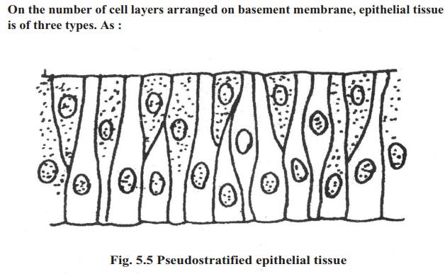
iii. Pseudo stratified Epithelial Tissue: .Cells, of this tissue are arranged in asingle layer on basement membrane. The cells are not of the same height. So this tissue appears to be stratified tissue (Fig-5.5).
Example: Trachea.
Besides the cells of epithelial tissue. are transformed variously for different functions. As:
1. Ciliated Epithelial Tissue: Found in the wall of the respiratory tube ofvertebrate animals.
2. Flagellated Epithelial Tissue: Found in the endoderm of Hydra.
3. Pseudopodia Epithelial Tissue: Found in pseudopodial cells in theendoderms of Hydra and in the intestine of vertebrate animals.
4. Glandular Epithelial Tissue: This is a kind of epithelial tissue transformedinto gland in the stomach and intestine of vertebrate animals.
5. Germinal Epithelial Tissue: This, is a kind of transformed epithelial tissue.From this tissue sperms and ovum are formed.
General Functions of Epithelial Tissue:
1. This tissue form, the external and internal- covering of any organ or tube.
2. After transformation this tissue takes part in , protection; secretion, absorption, diffusion, transportation etc. So, it can be said that epithelial tissue being transformed into glandular tissue and germinal tissue perform various important functions.
2 . Connective Tissue .
Structural Characteristics, Functions and Location of Connective Tissue .
Structural Characteristics: The. amount of matrix is more in connectivetissue but the number of cells is comparatively less. Matrix may be jelly like, soft or hard and fragile. One or more than one type of fibre and materials like calcium carbonate may remain present in matrix (Fig-5.6):
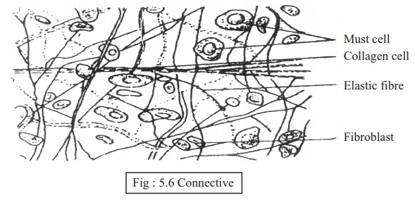
Functions: Connective tissue connects muscle with muscle and bone with bone. Connective tissue may transform into skeletal tissue, fibrous tissue and. fluid connective tissue.
On the basis of structure and function connective tissue is mainly of three types. As:
A. Films Connective Tissue: This type of connective tissue lies below thebody-integument and sparsely in muscles. In their matrix numerous fibres are present.
B. Skeletal Tissue: Internal structural building tissue of the body iscalled the skeletal tissue.
Functions:
1. This tissue forms the internal structure of the body, e.g. skeletal system.
2. It gives the body definite shape and firmness.
3. It helps in organ movement and locomotion.
4. I4. It protects the soft and sensitive organs of the body (as brain. spinal cord, lungs, heart etc.).
5. It produces various types of blood corpuscles.
6. It forms the surface for the attachment of voluntary muscles.
Depending on the formation, skeletat tissue is of two types.
a. Cartilage: Cartilage is a kind of flexible skeletal tissue. Their matrixes aresolid but they are soft and their cells have large spaces. Cartilage is suited at the two ends of the humerus, femur, and pinna of the ear and nose of the mammals.
b. Bone: Bone is hard, fragile and unflexible skeletal connective tissue.
Bones become rigid due to deposition of lime in their matrix. Some bones are solid. For an example, long bones of hind limb of vertebrates. Parts of long bones near-the bone cavities are sponge like.
c. Fluid Connective Tissue:
Structural Characteristics: Matrix of this tissue is liquid. In he matrix thereare various types of organic materials in the form of colloid.
Function: The main function of vascular tissue is to maintain circulation in theinterior of the body and resistance from disease. This tissue is of two types: Blood and Lymph.
1. Blood: Blood is a type of alkaline, stightly saline, red coloured, liquidconnective tissue. Flowing through the artery, veill and capillaries, blood takes part in internal circulation. Blood, blood vessels and heart together form circulatory system.
Structural Characteristics: Blood is formed of two components:
i. Plasma: It is the liquid part of blood. It is straw coloured. It contains(91-92) % water and (8-9) % organic and inorganic materials. The organic substances include various types of blood protein and wastematerials. The inorganic part contains different minerals like sodium, potassium, iron. calcium, magnesium etc.
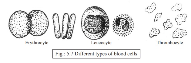
Blood Cell or Blood Corpuscles: Blood corpuscles form the majorcomponents of blood. Blood cells are of three types. These are:
Red Blood Corpuscle or Erythrocyte: These blood corpuscles containhaemoglobin. Due to haemoglobin colour of blood is red. the red blood corpuscles of the amphibians are biconvex, nucleated and oval. On the contrary, the red corpuscles of the blood of mammals are biconvex, non nucleated and round. Haemoglobin is combined with oxygen forms a unstable compound oxyhaemoglobin. It breaks down and releases oxygen in places where it is needed. '
Functions: To, carry oxygen and some carbon dioxide.
a. White Blood Corpuscle or Leucocyte: These generally lack definite shapeand are nucleated. Cytoplasm of white corpuscles are either granular or non granular.
Functions: To destroy germs and take part in self defense.
Thrombocyte or Platelets: These are present in the blood of vertebrateanimals. These are usually nucleated and spindle shaped. Nucleus is absent in the Thrombocytes of mammals. The thrombocyte of mammal is also called platelet.
Function:. Thrombocytes take part in blood coagulation or blood clotting.
2. Lymph:- The fluid materials stored in the spaces between different tissuesare collected by some small vessels. These small vessels are united together to form larger vessels. The system formed by these vessels known as lymphatic system. These vessels are lymph vessels and the' fluids are lymph. The large lymph vessels enter the vain in the shoulder region of man. There are some cells in the lymph known as lymphocyte. Lymph is a kind of slightly alkaline, transparent -and yellow coloured fluid.
3. The Muscular Tissue:
The particular type of contracting and expanding tissue originates from embryonic, mesoderm is called Muscular tissue (Fig-5.S).
Structural Characteristics: Matrix is nearly absent in muscular tissue. Themuscle cells are elongated and fibre-like. The fibres are spindle shaped. The fibres are known as myofibril. The cytoplasm of muscle cell may have one or more nuclei. The cell membrane of the muscle cell is known as sarcolemma. The myofibrils with transverse striation are known as striated muscle and without striation are known as smooth muscle.
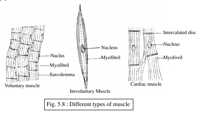
Function: Muscle cells through contraction or expansion take part in organmovement, locomotion and internal circulation. On the basis of location, structure and functions, muscle tissue is of three types. Such as:
Voluntary or Striated Muscle: This Type of muscular tissue can be contracted orexpanded at the will of the living beings. The cells of the voluntary muscle tissue are tubular, not branched and provided with transverse striations. These have generally more than one nucleus. This muscle can contract or expand quickly. This muscle is also, called striated or skeletal muscle.
Location: Voluntary muscles remain attached with the skeletal system as musclesof-hand, and feet of man.
Function: To control the movement and mobilization of different organs byvoluntary movement of various bones.
Involuntary or Smooth Muscle:
Structural Characteristics: The contraction and expansion of this muscle tissue isnot at all of the living beings. This muscle tissue is spindle shaped. Transverse striations are not present here. That is why this muscle is called smooth muscle.
Location: Involuntary muscles are found on the walls of blood vessels, alimentarycanal etc. of the vertebrate animals.
Function: Involuntary muscles mainly take part in the movement of internal organs,e.g. peristalsis. of intestine.
Cardiac Muscle :
Structural Characteristics: The special type of involuntary muscle that formsthe heart of vertebrate animals is called the cardiac muscle. The cells of this mucle tissue are tubular (very similar to those of voluntary muscle), branched and provided with transverse striations. Between the cells of this tissue intercalated disc are present. The contraction and relaxation of this tissue is not dependent on the will of the living beings. That is, the structure of heart muscles is ,like that of voluntary muscle and the function is like that of involuntary muscle. The cells of cardiac muscle, attached by branch joined together by branch. The contraction and expansion of all cardiac muscles take part combindly.
Function: Through rhythmic contraction and relaxation, the cardiac musclescontrol the circulation of blood within the body from a particular stage of the embryonic condition until death.
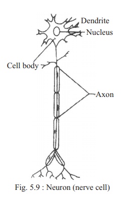
4. The Nerve Tissue:
Structural Characteristics: The particular type of tissue, which forms thenervous system, is called nerve tissue. Receiving stimulus from the environment, the nerve tissue transmits it within the body and accordingly appropriate responses are created. The special type of cell which forms the nerve tissue is called nerve cell or neuron (Fig-5.9) So a neuron is the structural and functional unit of nervous system. It is ectodermal in origin.
Neuron or nerve cells can receive various types of external and internal stimuli or nerve-sensation and can transmit those inside the body.
Structure of a Neuron: A mature neuron has three parts These are:
1. Cell body: The cell body is generallypolygonal and nucleated. The cytoplasm of the cell contains mitochondria, golgibody,ribosome, endoplasmic reticulum etc. But as there is no active centriole in the cytoplasm of neuron, the neuron cannot divide.
Function: Originating from the cell body, two or more branches transmitstimuli or nerve impulse to the neuron's cell body. Generally they are one or several in numbers and present opposite to the axon.
Axon : From the neuron's cell body a long fibre branch carries nerve impulsetowards dendrite of the next neuron. A neuron has only one axon. Between the with the dendrite of the other. It is called synapse. Through the synapse stimuli of a nerve is transmitted from one neuron to another.
Location : Nerve tissues are located within the nervous system. There are innumerable neurons is the nerve tissue of any animal.
Function :
1. To receive stimuli and create proper sensation.
2. To store memory in higher animals.
3. To control the works of different organs of the body and coordinate their activities.
Related Topics