Chapter: Diseases of The Brain and Nervous System(A Health Education Guide): Neuroradiology : Theimaging of The Brain
Neuroradiology - Theimaging of The Brain
NEURORADIOLOGY - THEIMAGING OF THE BRAIN
A doctor normally diagnoses and treats the patient through observations of symptoms, history etc. Many a times when it becomes necessary to know the location, extent and damage caused by a disease lying inside the body, radiology- neuroradiology becomes helpful and and necessary. Radiology-neuroradiology consists of Screening, X-Rays, Ultrasound, CT scan, and MRI etc.
X-Ray :
The “miraculous rays” X-Rays which could look-make structures inside the body were discovered in 1895 by a German scientist Mr: Roentegen. Thereafter these rays have been used in all possible innovative ways in the medical field. Sunrays, X-Rays, Microwave, Radio waves are all electro magnetic rays in scientific terminology. The only differentiating factor between them is their energy- content. Radio and television waves do not have much energy and so though they are present all around us in innumerable numbers, they cannot harm us and we are still alive. The energy from the X-Rays is 10,000 to 15,000 times more powerful than light and therefore can penetrate through objects. An amazing fact of nature is that the human eye can perceive only the sun rays and the rest of the rays are invisible to us.
The common X-Rays only give a rough picture of the skull or a one dimensional picture, but the depth of any feature cannot be judged, e.g. if there is a tumor inside thebrain its exact location as well as the depth of the abnormality cannot be judged by X-Rays.
C T Scan : (Computed Axial Tomography)
X-rays are used to give us a picture of different parts of the body. However, since we are now discussing about the diseases of the brain, it is necessary to know how X-Rays are helpful in this context. In the previous chapter we learned that the brain is well protected in the skull and therefore X-Rays are only able to give us the information of the skull. In-depth information regarding the inner structure cannot be obtained. The CT scan machine invented by a British Scientist Honsfield solved this problem. CT scan or CAT Scan is Computed Axial Tomography. CT scan also uses the X-Ray technology but with the help of the computer a three-dimensional view of the body is created. If there is a tumor in the brain, its depth and position can be determined by dividing the brain into imaginary parts like slices of bread, and each part is X-Rayed from various angles. The computer then calculates and creates a three- dimensional picture, which determines the depth and the size of the tumor very accurately. Thus, the minutest of changes inside the brain can also be determined with the help of the CT scan.
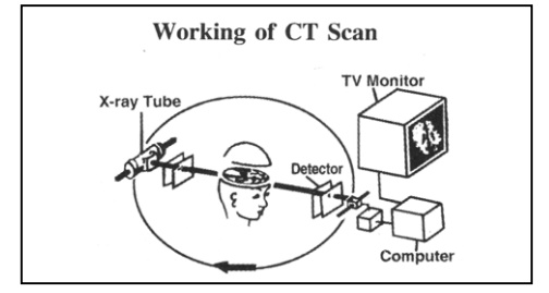
The diagram below explains how a photograph is taken with the help of a CT Scan.
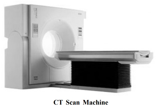
CT Scan Machine :
The CT Scan Machine is like a cubical box and is called Gantry. Within this box lies a two feet long circular tunnel. The stretcher like table on which the patient lies, can be moved in all directions upward, downward and side ways. The part of the body to be examined is kept at the center of the tunnel. The X-Ray tube lies in the middle of the tunnel, and rotates in a circular motion photographing the body part from each angle. These photographs are reflected on the detector, a computer carries out accurate calculations, divides the area into slices and then with the help of laser camera photographs are taken. The entire procedure takes about 15 to 30 minutes and during this time the patient has to lie still.
On an average twenty photographs are taken on a 14" × 17" X-Ray film, which are interpreted by the radiologist and a report is made. A CT scan can gives detailed informationof organs right from the brain to the spinal cord and lungs to the stomach, so it is necessary to write exactly which body part is to be scanned. The scanning procedure is painless and the patient just has to lie down still for a short period of time. In the diseases of brain like tumor or infection, a special drug called the Contrast Agent is injected intravenously to figure out the circulation of blood inside the diseased part. This iodine-based drug (Iodinised contrast) sometimes can make a person nauseate or temporarily increase his body temperature. However, the patient becomes normal within a short period of 1 to 2 minutes. Therefore, the patient is advised to fast for three hours prior to the contrast CT Scan. A patient suffering from allergy, asthma, kidney disorder or thyroid disease can suffer a reaction in contrast CT Scan, so in these cases, a non-ionic dye is used to prevent a reaction. A pregnant woman should inform the doctor of her condition prior to the examination. CT Scan has no side effects except those caused by radiation, so the relatives and friends of the patient are not allowed inside the CT Scan room and the rest of the body of the patient is kept covered.
CT Scan has now become a preliminary diagnostic procedure for the diseases of the Brain and is now available at all the cities of district level in India and abroad. The average expenses of a CT scan roughly range from Rs. 1700 to 2000.
The CT scan can offer only limited information about the structures like the white matter of the brain and the spinal cord etc. In 1972,a scientist called Damadian discovered the use of magnetic field in the examination of the human body. Advanced computers are used to photograph the brain by using powerful magnetic fields. This is known as the Magnetic Resonance Imaging (M.R.L). MRI has proved very beneficial in the examination and diagnosis of diseases like the brain tumor, paralysis, white matter disease, congenital disorders, and disease of the nervous system e.g. multiple sclerosis and the examination of the minutest and innermost parts of eyes and ears.
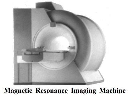
MRI Machine :
The MRI machine is cylindrical in shape, made of a powerful magnet. The patient lies in the tunnel formed in this cylinder. This magnet is 1000 times more powerful than the magnetic field of the earth. The power of the machine is determined through a measuring unit called Tesla. Normally the machine is configured as 0.2T, 0.3T, 0.5T, LOT and 1.5T. As the Tesla (power) increases, the accuracy, speed and minuteness of the machine also increase. Superior quality of machines use Helium gas to maintain the magnetic field and therefore the MRI machine has to be placed in an air-conditioned room.
Like C T Scan, MRI can also be used for the examination of organs ranging from the brain to spinal cord, stomach, kidney, lungs, bones, muscles etc. The patient is positioned in such a way that the organ to be examined remains at the centre of the machine. Here the X-Rays are not used, instead radio frequency waves and strong magnetic field are used, this eliminates the fear of harmful radiation. Hydrogen protons are present in the water, which are present in a large quantity in the body. According to the principles of MRI, radio frequency waves and magnetic waves excite these protons. Each cell has a different number of protons and with the help of radio signals and advanced computers one can accurately calculate the various numbers of protons and thus differentiate each and every type of cell which can be photographed from every angle, with the help of a laser camera on a 14" x 17" photo film. This can easily differentiate between the white and the grey matter of the brain. On a photo plate roughly 16 to 20 photographs can be taken and the entire MRI consists of 4 to 5 such sequences. Each sequence lasts for about 5 to 8 minutes. Thus, in 30 to 45 minutes, 80 to 100 photographs of the brain from the different angles (X, Y, Z, axis) can be taken, which an expert radiologist analyzes and a report is given. This can be considered as an amazing gift of modern medical science to mankind.
During MRI test, the gradients of the machine make a lot of sound The sound can be muffled by using cotton balls, earplugs or earphone. Sometimes music is also played in the room. Since the patient has to lie down in an enclosed cylinder, some of them may feel claustrophobic. Such patients may be given mild anesthesia. Latest machines have an open magnet and the machine structure is open and so the patientsfeel less claustrophobic. In some diseases, a special drug (contrast) has to be used to specify some defects; this drug has very few side effects.
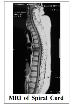
The MRI scan is much more advanced compared to the CT scan. First of all, an X-Ray is not used in MRI and so it is far less risky than CT scan during pregnancy. Just as we have seen earlier, in CT scan only bread slice cuts (axial) can be seen, while in MRI one gets a three dimensional (X, Y, Z) view; hence a complete picture is seen. The spinal cord can be minutely seen only by an MRI. Because of the strong magnetic field, things like watch, credit card, pen, rings etc. are not permitted in the MRI room. Patients who have a pace maker in their heart or any other electronic gadget in their body cannot undergo MRI. New research has made diffusion and perfusion MRI possible. This makes the diagnosis of hampered blood circulation in any part of the brain in minutes, so prompt treatment can prevent disease like paralysis.
Functional MRI is a discovery that can differentiate various areas of brain associated with movement, memory, speech, emotions etc. The major benefit of this discovery is in the planning of surgery and radiotherapy. The important centers in the brain can be avoided during surgery and the patient can be saved from permanent disability.
The cost of MRI is roughly Rs.5000 to 7000 and this facility is now available in all major cities of India.
Angiography is the examination of the arteries and veins that carry blood to the various parts of the body. Now-a-days, even a layman is also aware of the angiography of the heart. The blood vessels of the brain are also investigated in a similar mariner.

The Digital Subtraction Angiography (DSA) is used for carrying out the angiography of the brain in many diseases of the brain, like constriction of blood vessels, arteriosclerosis, aneurysms, AV malformation etc. In thisprocedure a catheter is introduced from the vein in the thigh. The catheter is pushed inside along with the blood flow. With the help of X-Ray and the computer monitor, the catheter is made to enter the vessels of the brain. After that a special drug (contrast medium) is injected. As the drug enters the bloodstream, its progression in the blood vessels is seen live on a monitor and if necessary, with the help of X-Rays the blood vessels are also visualized from various angles. In Digital Subtraction Angiography, an X-Ray of the brain is taken before starting the procedure. Another X-Ray is taken after the introduction of the contrast medium and the differences in the vessels and brain seen in both the X-Rays are used for diagnosis. If the Lumen of the carotid artery, which passes across the neck and supplies blood to the brain, becomes narrow due to arteriosclerosis, it can be widened with the help of a balloon. This is known as carotid angioplasty.
The risk factor of the angiography of the brain is similar to the angiography of the heart. Certain nominal risks are very much there, but they can be tackled. The investigation of the blood vessels is possible by totally non-risky methods too, but the information gained by them is 5-10% less accurate. These methods include, Color Doppler, CT Angiography, and MRI Angiography etc. Color Doppler gives complete information regarding the Carotid artery. The diameter of the Artery, the pressure flow of the blood and the deposits in the wall can be determined easily using simple Sonography. MRI Angiography can minutely examine the arteries and veins without the use of a catheter and is now exceedingly used as a primary investigative tool for blood vessels. As in MRI, this investigation also requiresfor the patient to simply lie down on the investigation table. Science, technology and the modern computers have brought innovative advancements in the field of radiology at a very fast pace. PET SCAN (Positron Emission Tomography) can give much advanced information regarding certain diseases and can be used for better treatment of the patient. However this diagnostic tool is not easily available. But the use of the SPECT Scan for the investigation of the efficiency and metabolism of the brain is becoming increasingly rampant.
The invention of the radiation and the X-rays is not only important in the field of health, but is useful in other sectors too. Till now, 15 Nobel prizes have been awarded to various inventors doing researches using X-Rays. This can be considered a parameter to determine the extent to which X-rays are useful in human life. More research and new inventions are still taking place in this field. We hope that the advancement of this technology gives maximum health benefits to mankind.
Related Topics