Chapter: Medical Physiology: Cerebral Blood Flow, Cerebrospinal Fluid, and Brain Metabolism
Cerebrospinal Fluid Pressure
Cerebrospinal Fluid Pressure
The normal pressure in the cerebrospinal fluid system when one is lying in a horizontal position averages 130 millimeters of water (10 mm Hg), although this may be as low as 65 millimeters of water or as high as 195 mil-limeters of water even in the normal healthy person.
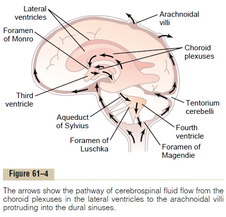
Regulation of Cerebrospinal Fluid Pressure by the Arachnoidal Villi. The normal rate of cerebrospinal fluid formationremains very nearly constant, so that changes in fluid formation are seldom a factor in pressure control. Con-versely, the arachnoidal villi function like “valves” that allow cerebrospinal fluid and its contents to flow readily into the blood of the venous sinuses while not allowing blood to flow backward in the opposite direction. Nor-mally, this valve action of the villi allows cerebrospinal fluid to begin to flow into the blood when cerebrospinal fluid pressure is about 1.5 mm Hg greater than the pres-sure of the blood in the venous sinuses. Then, if the cere-brospinal fluid pressure rises still higher, the valves open more widely, so that under normal conditions, the cere-brospinal fluid pressure almost never rises more than a few millimeters of mercury higher than the pressure in the cerebral venous sinuses.
Conversely, in disease states, the villi sometimes become blocked by large particulate matter, by fibrosis, or by excesses of blood cells that have leaked into the cerebrospinal fluid in brain diseases. Such blockage can cause high cerebrospinal fluid pressure, as follows.
High Cerebrospinal Fluid Pressure in Pathological Conditions of the Brain. Often a largebrain tumorelevates the cere-brospinal fluid pressure by decreasing reabsorption of the cerebrospinal fluid back into the blood. As a result, the cerebrospinal fluid pressure can rise to as much as 500 millimeters of water (37 mm Hg) or about four times normal.
The cerebrospinal fluid pressure also rises consider-ably when hemorrhage or infection occurs in the cranial vault. In both these conditions, large numbers of red and/or white blood cells suddenly appear in the cere-brospinal fluid, and they can cause serious blockage of the small absorption channels through the arachnoidal villi. This also sometimes elevates the cerebrospinal fluid pressure to 400 to 600 millimeters of water (about four times normal).
Some babies are born with high cerebrospinal fluid pressure. This is often caused by abnormally high resist-ance to fluid reabsorption through the arachnoidal villi, resulting either from too few arachnoidal villi or from villi with abnormal absorptive properties. This is dis-cussed later in connection with hydrocephalus.
Measurement of Cerebrospinal Fluid Pressure. The usual pro-cedure for measuring cerebrospinal fluid pressure is very simple and is the following: First, the person lies exactly horizontally on his or her side so that the fluid pressure in the spinal canal is equal to the pressure in the cranial vault. A spinal needle is then inserted into the lumbar spinal canal below the lower end of the cord, and the needle is connected to a vertical glass tube that is open to the air at its top. The spinal fluid is allowed to rise in the tube as high as it will. If it rises to a level 136 millimeters above the level of the needle, the pres-sure is said to be 136 millimeters of water pressure or, dividing this by 13.6, which is the specific gravity of mercury, about 10 mm Hg pressure.
High Cerebrospinal Fluid Pressure Causes Edema of the Optic Disc—Papilledema. Anatomically, the dura of the brainextends as a sheath around the optic nerve and then connects with the sclera of the eye. When the pressure rises in the cerebrospinal fluid system, it also rises inside the optic nerve sheath. The retinal artery and vein pierce this sheath a few millimeters behind the eye and then pass along with the optic nerve fibers into the eye itself. Therefore, (1) high cerebrospinal fluid pressure pushes fluid first into the optic nerve sheath and then along the spaces between the optic nerve fibers to the interior of the eyeball; (2) the high pressure decreases outward fluid flow in the optic nerves, causing accumu-lation of excess fluid in the optic disc at the center of the retina; and (3) the pressure in the sheath also impedes flow of blood in the retinal vein, thereby increasing the retinal capillary pressure throughout the eye, which results in still more retinal edema.
The tissues of the optic disc are much more distensi-ble than those of the remainder of the retina, so that the disc becomes far more edematous than the remainder of the retina and swells into the cavity of the eye. The swelling of the disc can be observed with an ophthal-moscope and is called papilledema. Neurologists can estimate the cerebrospinal fluid pressure by assessing the extent to which the edematous optic disc protrudes into the eyeball.


Related Topics