Chapter: Pathology: Hematopoetic Pathology–White Blood Cell Disorders & Lymphoid and Myeloid Neoplasms
Reactive Changes in White Blood Cells
REACTIVE CHANGES IN WHITE BLOOD CELLS
Leukocytosis
Leukocytosis is characterized by
an elevated white blood cell count. It has the fol-lowing features:
•
Increased neutrophils (neutrophilia)
Increased bone marrow production
is seen with acute inflammation associated with pyogenic bacterial infection or
tissue necrosis
Increased release from bone
marrow storage pool may be caused by corti costeroids, stress, or endotoxin
Increased bands (“left shift”)
noted in peripheral circulation
Reactive changes include Döhle
bodies (aggregates of rough endoplasmic reticulum), toxic granulations
(prominent granules), and cytoplasmic vacuoles of neutrophils
•
Increased eosinophils (eosinophilia) occurs with allergies and asthma
(type I hypersensitivity reaction), parasites, drugs (especially in hospitals),
and certain skin diseases and cancers (adenocarcinomas, Hodgkin disease).
•
Increased monocytes (monocytosis) occurs with certain chronic diseases
such as some collagen vascular diseases and inflammatory bowel disease, and
with certain infections, especially TB.
•
Increased lymphocytes (lymphocytosis) occurs with acute (viral) diseases
and chronic inflammatory processes.
Infectious mononucleosis, an
acute, self-limited disease, which usually resolves in 4–6 weeks, is an example
of a viral disease that causes lymphocytosis. The most common cause is
Epstein-Barr virus (a herpesvirus) though other viruses can cause it as well (heterophile-negative
infectious mononucleosis is most likely due to cytomegalovirus).
Age groups include adolescents
and young adults (“kissing disease”).
The “classic triad” includes
fever, sore throat with gray-white membrane on tonsils, and lymphadenitis
involving the posterior auricular nodes. Another sign is hepatosplenomegaly.
Complications include hepatic
dysfunction, splenic rupture, and rash if treated with ampicillin.
Diagnosis is often made based on
symptoms. Lymphocytosis and a rising titer of EBV antibodies are suggestive of
the infection. Atypical lymphocytes may be present in peripheral blood.
Mono-spot test is often negative early in infection.
•
Increased basophils are seen with chronic myeloproliferative disorders
such as polycythemia vera.
Leukopenia
Leukopenia is characterized by a
decreased white blood cell count. It has the fol-lowing features:
•
Decreased neutrophils can be due to decreased production (aplastic
anemia, chemotherapy), increased destruction (infections, autoimmune disease such
as systemic lupus erythematosus), and activation of neutrophil adhesion
mol-ecules on endothelium (as by endotoxins in septic shock).
•
Decreased eosinophils are seen with increased cortisol, which causes
seques-tering of eosinophils in lymph nodes; examples include Cushing syndrome
and exogenous corticosteroids.
•
Decreased lymphocytes are seen with immunodeficiency syndromes such as
HIV, DiGeorge syndrome (T-cell deficiency), and severe combined
immu-nodeficiency (B- and T-cell deficiency); also seen secondary to immune
destruction (systemic lupus erythematosus), corticosteroids, and radiation
(lymphocytes are the most sensitive cells to radiation).

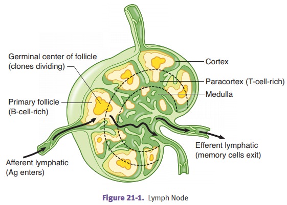
Lymphadenopathy
Lymphadenopathy is lymph node enlargement due
to reactive conditions or neo-plasia.
Acute nonspecific lymphadenitis produces tender enlargement
of lymph nodes; focal involvement is seen with bacterial
lymphadenitis. Microscopi-cally, there may be neutrophils within the lymph
node. Cat scratch fever (due to Bartonella
henselae) causes stellate microabscesses. Generalized involve-ment of lymph
nodes is seen with viral infections.
Chronic nonspecific lymphadenitis causes nontender enlargement
of lymph nodes. Follicular hyperplasia involves B lymphocytes
and may be seen with rheumatoid arthritis, toxoplasmosis, and early HIV
infections. Paracortical lymphoid hyperplasia involves T cells and may be seen
with viruses, drugs (Dilantin), and systemic lupus erythematosus. Sinus
histiocytosis involves macrophages and, in most cases, is nonspecific; an
example is lymph nodes draining cancers.
Neoplasia usually causes nontender enlargement of lymph
nodes. The most common tumor to involve lymph
nodes is metastatic cancer (e.g., breast, lung, malignant melanoma, stomach and
colon carcinoma), which is initially seen under the lymph node capsule. Other
important causes of lymphadenopathy are malignant lymphoma and infiltration by
leukemias.
Related Topics