Chapter: Diseases of The Brain and Nervous System(A Health Education Guide): Brain Hemorrhage
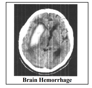
Brain Hemorrhage

BRAIN HEMORRHAGE
Brain Hemorrhage is a more serious type of brain attack and accounts for 20% of the cases of strokes occurring due to the faults in blood vessels. The remaining occur due to thromboembolism. This is a serious disease of the brain in which there is bleeding in the brain either due to the rupture of a blood vessel or some other reason. Most of the patients become unconscious in minutes and if timely treatment is not given, it proves fatal for many patients.
Brain hemorrhage can be classified into two groups:
1. Intracerebral Hemorrhage: Occurs due to either high blood pressure or because of the accumulation of a substance called Amyloid (Amyloid Angiopathy) in the blood vessel.
2. Subarachnoid Hemorrhage: an unnatural swelling on the blood vessel (saccular aneurysm), or formation ofan entangled mass of abnormal blood vessels (AN. Malformation) might rupture and cause hemorrhage.
In addition to these, side effects of anticoagulant type of medicines, head injuries, disorders of coagulation, infection in the brain, rupture of cancerous tumors can also cause brain hemorrhage.
1. Intra Cerebral Hemorrhage :
Rupture of blood vessels deep inside the brain due to high blood pressure is called intracerebral hemorrhage. This hemorrhage occurs at some particular locations in the brain (like Putamen, Thalamus, Cerebellum) and usually while examining the patient; the physician can easily identify the location, by its specific signs and symptoms.
Amyloid Angiopathy is s a kind of cerebral hemorrhage occurring mostly in elderly people and it can recur frequently.
If all these hemorrhages are diagnosed quickly and immediate treatment is initiated to reduce the edema of the brain and control of blood pressure, the death rate in the cases of cerebral hemorrhage can be brought down considerably. At present the death rate is as high as 50% to 60%. In some cases of cerebellarhemorrhage, e.g. temporal lobe hemorrhage or putaminal hemorrhage, lives can be saved by surgery done by a neurosurgeon. The goal of reducing death rate due to cerebral hemorrhage can be achieved by factors like awareness about the disease, quick diagnosis, treatment at a war footing, expert and quick decision making physicians and neurosurgeons and hospitals with all amenities like ventilator machine, operation theatre etc.
Symptoms :
Sudden headache, vomiting, vertigo, blackouts (these can be due to high blood pressure also), seizures, stumbling, paralysis, and loss of consciousness within a few minutes with rapid breathing; are the usual symptoms of brain hemorrhage.
Diagnosis and emergency treatment :
It is essential to get an immediate diagnosis with a CT scan or MRI scan. These tests can also identify the location of the hemorrhage, the size of the clot, edema of the brain and the cause of the same. It is better to get a CT scan done even before admission, if the facility is available in the city or town, provided the patient is stable with normal respiration and B.P.. This is for confirmation of the hemorrhage. Even if clinically, a hemorrhage is suspected, the scan some times may reveal a thrombosis, tumor, subdural or cerebral infection’ and that would make a major difference in the line of treatment. However, emergency treatment in a hospital should be given immediately and the scan may be done later if the condition of the patient is serious.
It is essential to create awareness in the public that in any serious neurological condition, instead of wasting time by insisting on a home visit by the specialist, it is advisable to immediately call the family doctor and rush the patient to the hospital with an ambulance and if required get a scan done before admission. If the specialist doctor can visit immediately, it would be excellent. But usually it may take 2 to 4 hours for him to be available, during which precious time is lost and the delay in treatment may cause irreparable damage to the brain. If the family doctor also is unable to make it on time,then the best option would be to take the patient immediately to the emergency ward of a good hospital and arrange for the specialist to reach there. Such a detailed explanation has basically been given on this subject because in majority of the cases exactly the opposite is seen to be happening and it leads to immense regret. Emergency medicine and, critical care is a separate and extremely important aspect of the medical fraternity. Here every second counts and critical decisions taken by the specialized doctors, who are well trained to save lives, play a very important role. Therefore, in this matter no social arguments or interference should be entertained.
In a case of hemorrhage, if blood pressure is found high by the family physician, immediate treatment for controlling the blood pressure is given. If there is an indication of thrombosis, then the blood pressure should not be abruptly brought down as it can cause a lot of damage. But if the blood pressure is very high or the patient is suffering from heart disease, or the patient is on anticoagulant therapy, it becomes very essential to lower the blood pressure to normal level even in case of thrombosis.
If the edema of the brain seems substantial, then emergency injections (mannitol, lasix) can be given by the family physician at home, before transferring the patient to the hospital. If the patient is getting seizures, then one should not wait, but start urgent treatment at home.
Sometimes lumbar puncture could be useful apart from CT scan, in the diagnostic process, but as mentioned later in the chapter on brain tumors, lumbar puncture is to be avoided if there is edema in the brain. Here it may prove dangerous.Naturally, these patients are kept in ICU They are monitored very intensively along with emergency treatment: If required angiography is performed and in selected cases surgery is done to aspirate blood.
If the cause of the hemorrhage is a deficiency of any of the blood clotting factors, the deficiency is corrected by transfusion of those factors. If the hemorrhage has occurred due to the side effects of any drug (like Warf, Acetrom which are given in cases of valvular defects) then plasma and other appropriate blood components are transfused to stop hemorrhage. In the medical profession hemorrhage is one of the most serious side effects of these drugs. The drugs, which prevent the clotting of the blood, can -cause hemorrhage due to overdose in some cases. Therefore, it is very important to inform the patient in detail about the side effects of the medicine. Every 7 to 15 days Prothrombin time APTT/INR blood test has to be carried out regularly to monitor the thinning of the blood. If proper precautions are taken, no side effects occur and the patients lead a complication free life for years at a stretch. Just as insulin is extremely beneficial for a diabetic patient to lead a normal life, but an unrequired higher dose may cause hypoglycemia and even death. Same holds true for anticoagulant drugs.
Thus regulation of blood pressure, medicines for the edema of the brain, proper nursing, treatment of complications, and if required surgery can save the patients of intra cerebral hemorrhage to a large extent. If the patient is saved but paralysis persists as a consequence of the hemorrhage, physiotherapy along with drugs should be used for a longperiod of time to activate the paralyzed limbs once again. It is a fact that the initial chances of death are much higher in hemorrhage than in thrombosis, but so is also a fact that recovery from paralysis due to hemorrhage is much better than in thrombosis. For care of sick patients, certain guidelines are given in chapter 24, which may be followed strictly by the care taker.
2. Subarachnoid Hemorrhage :
This type of hemorrhage is completely different from the one discussed before. Most of these patients do not suffer from high blood pressure. Many of them are young and in most of them there is a congenital weakness in the blood vessels causing ballooning of the blood vessel (Saccular aneurysm) or entanglement of the vessels (AN. Malformation). These which rupture at a particular age due to sudden exertion or unknown causes, with oozing of blood into the subarachnoid spaces between the membranes of the brain. This is known as the subarachnoid hemorrhage. It is worth noting that out of every 100 people, at least one may be harboring such a congenital aneurysm in the blood vessels of the brain, but it can never be predicted when it may rupture and in many cases it may not rupture during the entire course of life.
Once it ruptures, nearly 45% to 60% patients die within a month. This disease is extremely dangerous and hence it is not only important to understand the disease but also have its early diagnosis before it ruptures. It is surprising that many patients may suddenly suffer from hemorrhage without any warning symptoms. However according to a group ofspecialists, if there is a migraine like headache, strictly only on one side of the brain (like right side, above the ear, behind the eyes), which does not shift to the other side, and recurs frequently, an MR angiography of the brain should be done as a precautionary measure. This test can detect aneurysms of the blood vessels of the brain with an accuracy of 95 to 98%, without any invasive procedure. The cost of this test is about Rs.4000 to 6000/- but whether this test should be done in all such cases is debatable. But in my personal opinion and experience, in migraines, which constantly affect only one particular side of the brain, it is better to get this test done, to exclude aneurysm or A Vmalformation.
The main symptoms of this disease i.e. subarachnoithemorrhage are that the patient experiences a never-before kind of severe headache, sometimes accompanied by a seizure and he might even become unconscious momentarily due to the edema in the brain. Usually the patient regains consciousness in a short while, but may again start losing consciousness after some time, suffer from paralysis and there may be irregularities in the vital functions like respiration, blood pressure or heart. Many such patients die immediately or within 14 to 30 days. Therefore, if the patient feels that he/she has never experienced such a splitting headache before along with other signs and symptoms, it is all the more important for the patient to see a neurologist so that timely treatment may save his / her life.
Investigation of Blood Vessels of the Brain :
The test known as Angiography is the most important test. MR Angiography makes the use of the MRI magnetfor the examination of the blood vessels In this test no catheter is required to be introduced in the blood vessels. Therefore, it is called a Non Invasive test. For coronary angiography, a catheter has to be introduced in a blood vessel and therefore there is a slight risk involved, which is not the case in a MR Angiography. This test can give an accuracy of only 90 to 95% and therefore, can be used only as a screening test. The gold standard tests are the conventional 4 vessel angiography or Digital Substraction Angiography (DSA). An aneurysm is immediately detected on angiography. In 15% cases more than one aneurysm can be present and so it is imperative that the angiography is done on all the four blood vessels of the brain, so that if surgery becomes necessary it can be planned keeping all the aneurysms in mind.
The angiography can also detect another vascular condition, i:e the entanglement of blood vessels known as arteriovenous malformation (AN. Malformation). These patients have a history of severe headache; with an occasional seizure, and some of them may..already be suffering from the paralysis of a limb.
In some cases of sub-arachnoid hemorrhage DSA angiography may show normal results. Out of these cases a few may have a very small aneurysm or a cryptic AN. malformation or a vasospasm, which may not be detected on angiography. Therefore in such cases angiography is repeated after 3 months. Only then it can be confirmed that these are not the causes of hemorrhage. This kind of hemorrhage is termed as Idiopathic Sub-arachnoid Hemorrhage.
Complications :
In this disease, there is a definite risk of rebleeding due to rerupture of the aneurysm, any time in the next month. Usually this is fatal or very serious. Similarly, between 4 to 12 days of primary bleeding, there is a spasm in the vessel distal to aneurysm - so called vasospasm. This is the cause of delayed paralysis and morbidity. This can be largely prevented with the help of drugs and hydration. Further, salt depletion and seizures are also nons known complications.
So, once the diagnosis is confirmed, in appropriate cases the aneurysm is clipped by surgery. It is either covered by a tissue or a plastic coating is done. Sometimes the Carotid artery in the neck is tied for the treatment. In the cases of large aneurysms, treatment is also done with the help of balloon.
For treating AV Malformation, Block resection or Ligation Technique is used or it can be cauterized by a proton beam. Now-a-days gamma knife is more frequently used where gamma rays generated from a cobalt source are focused by a gamma knife and the cauterization is done with precision.
In those cases where surgery is not possible, embolisation is carried out by platinum foil. This technique can be used both in aneurysm as well as in AV Malformation. Luckily, all these surgical techniques and procedures are available in major cities in India and hence the mortality and morbidity are reduced very significantly.
Related Topics