Chapter: Medical Physiology: Behavioral and Motivational Mechanisms of the Brain-The Limbic System and the Hypothalamus
Hypothalamus, a Major Control Headquarters for the Limbic System
Hypothalamus, a Major Control Headquarters for the Limbic System
The hypothalamus, despite its very small size of only a few cubic centimeters, has two-way communicating pathways with all levels of the limbic system. In turn, it and its closely allied structures send output signals in three directions: (1) backward and downward to the brain stem, mainly into the reticular areas of the mes-encephalon, pons, and medulla and from these areas into the peripheral nerves of the autonomic nervous system; (2) upward toward many higher areas of the diencephalon and cerebrum, especially to the anterior thalamus and limbic portions of the cerebral cortex; and (3) into the hypothalamic infundibulum to control or partially control most of the secretory functions of both the posterior and the anterior pituitary glands.
Thus, the hypothalamus, which represents less than 1 per cent of the brain mass, is one of the most impor-tant of the control pathways of the limbic system. It controls most of the vegetative and endocrine func-tions of the body as well as many aspects of emotional behavior. Let us discuss first the vegetative and endocrine control functions and then return to the behavioral functions of the hypothalamus to see how these operate together.
Vegetative and Endocrine Control Functions of the Hypothalamus
The different hypothalamic mechanisms for control-ling multiple functions of the body are so important. To illustrate the organization of the hypothalamus as a functional unit, let us summarize the more important of its vegetative and endocrine functions here as well.
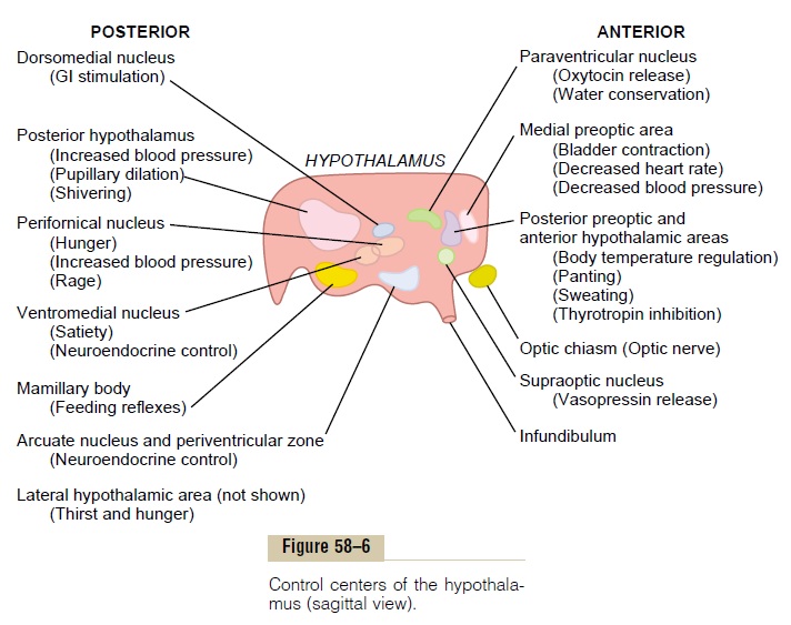
Figures 58–6 and 58–7 show enlarged sagittal and coronal views of the hypothalamus, which represents only a small area in Figure 58–4. Take a few minutes to study these diagrams, especially to see in Figure 58–6 the multiple activities that are excited or inhib-ited when respective hypothalamic nuclei are stimu-lated. In addition to the centers shown in Figure 58–6, a large lateral hypothalamic area (shown in Figure 58–7) is present on each side of the hypothalamus. The lateral areas are especially important in controlling thirst, hunger, and many of the emotional drives.
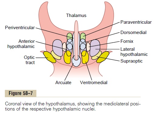
A word of caution must be issued for studying these diagrams because the areas that cause specific activi-ties are not nearly as accurately localized as suggested in the figures. Also, it is not known whether the effects noted in the figures result from stimulation of specific control nuclei or whether they result merely from activation of fiber tracts leading from or to control nuclei located elsewhere. With this caution in mind, we can give the following general description of the veg-etative and control functions of the hypothalamus.
Cardiovascular Regulation. Stimulation of different areasthroughout the hypothalamus can cause every known type of neurogenic effect on the cardiovascular system, including increased arterial pressure, decreased arterial pressure, increased heart rate, and decreased heart rate. In general, stimulation in the posterior and lateral hypo-thalamus increases the arterial pressure and heart rate,whereas stimulation in the preoptic area often has oppo-site effects, causing a decrease in both heart rate and arterial pressure. These effects are transmitted mainly through specific cardiovascular control centers in the reticular regions of the pons and medulla.
Regulation of Body Temperature. The anterior portion of thehypothalamus, especially the preoptic area, is concerned with regulation of body temperature. An increase in the temperature of the blood flowing through this area increases the activity of temperature-sensitive neurons, while a decrease in temperature decreases their activ-ity. In turn, these neurons control mechanisms for increasing or decreasing body temperature.
Regulation of Body Water. The hypothalamus regulatesbody water in two ways: (1) by creating the sensation of thirst, which makes the animal or person drink water, and (2) by controlling the excretion of water into the urine. An area called the thirst center is located in the lateral hypothalamus. When the fluid electrolytes in either this center or closely allied areas become too concentrated, the animal develops an intense desire to drink water; it will search out the nearest source of water and drink enough to return the electrolyte con-centration of the thirst center to normal.
Control of renal excretion of water is vested mainly in the supraoptic nuclei. When the body fluids become too concentrated, the neurons of these areas become stimulated. Nerve fibers from these neurons project downward through the infundibulum of the hypothala-mus into the posterior pituitary gland, where the nerve endings secrete the hormone antidiuretic hormone (also called vasopressin). This hormone is then absorbed into the blood and transported to the kidneys where it acts on the collecting ducts of the kidneys to cause increased reabsorption of water. This decreases loss of water into the urine but allows continuing excretion of electrolytes, thus decreasing the concentration of the body fluids back toward normal.
Regulation of Uterine Contractility and of Milk Ejection from the Breasts. Stimulation of theparaventricular nucleicausestheir neuronal cells to secrete the hormone oxytocin. This in turn causes increased contractility of the uterus as well as contraction of the myoepithelial cells sur-rounding the alveoli of the breasts, which then causes the alveoli to empty their milk through the nipples.
At the end of pregnancy, especially large quantities of oxytocin are secreted, and this secretion helps to promote labor contractions that expel the baby. Then, whenever the baby suckles the mother’s breast, a reflex signal from the nipple to the posterior hypothalamus also causes oxytocin release, and the oxytocin now per-forms the necessary function of contracting the ductules of the breast, thereby expelling milk through the nipples so that the baby can nourish itself.
Gastrointestinal and Feeding Regulation. Stimulation ofseveral areas of the hypothalamus causes an animal to experience extreme hunger, a voracious appetite, and an intense desire to search for food. One area associated with hunger is the lateral hypothalamic area. Con-versely, damage to this area on both sides of the hypo-thalamus causes the animal to lose desire for food, sometimes causing lethal starvation.
A center that opposes the desire for food, called the satiety center, is located in the ventromedial nuclei. When this center is stimulated electrically, an animal that is eating food suddenly stops eating and shows complete indifference to food. However, if this area is destroyed bilaterally, the animal cannot be satiated; instead, its hypothalamic hunger centers become overactive, so that it has a voracious appetite, resulting eventually in tremendous obesity. Another area of the hypothalamus that enters into overall control of gas-trointestinal activity is the mamillary bodies; these control at least partially the patterns of many feeding reflexes, such as licking the lips and swallowing.
Hypothalamic Control of Endocrine Hormone Secretion by the Anterior Pituitary Gland. Stimulation of certain areas of thehypothalamus also causes theanterior pituitary gland to secrete its endocrine hormones. This subject is discussed in detail in relation to neural control of the endocrine glands. Briefly, the basic mechanisms are the following.
The anterior pituitary gland receives its blood supply mainly from blood that flows first through the lower part of the hypothalamus and then through the anterior pituitary vascular sinuses. As the blood courses through the hypothalamus before reaching the anterior pitu-itary, specific releasing andinhibitory hormones are secreted into the blood by various hypothalamic nuclei. These hormones are then transported via the blood to the anterior pituitary gland, where they act on the glandular cells to control release of specific anterior pituitary hormones.
Summary. A number of areas of the hypothalamuscontrol specific vegetative and endocrine functions. These areas are still poorly delimited, so much so that the specification given above of different areas for dif-ferent hypothalamic functions is partially tentative.
Behavioral Functions of the Hypothalamus and Associated Limbic Structures
Effects Caused by Stimulation. In addition to the vegeta-tive and endocrine functions of the hypothalamus, stimulation of or lesions in the hypothalamus often have profound effects on emotional behavior of animals and human beings.
In animals, some of the behavioral effects of stimu-lation are the following:
1.Stimulation in the lateral hypothalamus not only causes thirst and eating, as discussed above, but also increases the general level of activity of the animal, sometimes leading to overt rage and fighting, as discussed subsequently.
2.Stimulation in the ventromedial nucleus and surrounding areas mainly causes effects opposite to those caused by lateral hypothalamic stimulation—that is, a sense of satiety, decreasedeating, and tranquility.
3.Stimulation of a thin zone of periventricularnuclei, located immediately adjacent to the thirdventricle (or also stimulation of the central gray area of the mesencephalon that is continuous with this portion of the hypothalamus), usually leads to fear and punishment reactions.
4.Sexual drive can be stimulated from severalareas of the hypothalamus, especially the most anterior and most posterior portions of the hypothalamus.
Effects Caused by Hypothalamic Lesions. Lesions in thehypothalamus, in general, cause effects opposite to those caused by stimulation. For instance:
1.Bilateral lesions in the lateral hypothalamus will decrease drinking and eating almost to zero, often leading to lethal starvation. These lesions cause extreme passivity of the animal as well, with loss of most of its overt drives.
2.Bilateral lesions of the ventromedial areas of the hypothalamus cause effects that are mainly opposite to those caused by lesions of the lateral hypothalamus: excessive drinking and eating as well as hyperactivity and often continuous savagery along with frequent bouts of extreme rage on the slightest provocation.
Stimulation or lesions in other regions of the limbic system, especially in the amygdala, the septal area, and areas in the mesencephalon, often cause effects similar to those elicited from the hypothalamus. We will discuss some of these in more detail later.
“Reward” and “Punishment” Function of the Limbic System
From the discussion thus far, it is already clear that several limbic structures are particularly concerned with the affective nature of sensory sensations—that is, whether the sensations are pleasant or unpleasant. These affective qualities are also called reward or pun-ishment, or satisfaction oraversion. Electrical stimula-tion of certain limbic areas pleases or satisfies the animal, whereas electrical stimulation of other regions causes terror, pain, fear, defense, escape reactions, and all the other elements of punishment. The degrees of stimulation of these two oppositely responding systems greatly affect the behavior of the animal.
Reward Centers
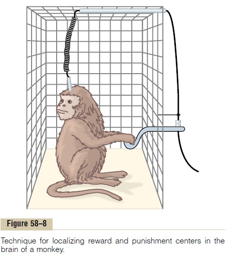
Figure 58–8 shows a technique that has been used for localizing specific reward and punishment areas of the brain. In this figure, a lever is placed at the side of the cage and is arranged so that depressing the lever makes electrical contact with a stimulator. Electrodes are placed successively at different areas in the brain so that the animal can stimulate the area by pressing the lever. If stimulating the particular area gives the animal a sense of reward, then it will press the lever again and again, sometimes as much as hundreds or even thousands of times per hour. Furthermore, when offered the choice of eating some delectable food as opposed to the opportunity to stimulate the reward center, the animal often chooses the electrical stimulation.
By using this procedure, the major reward centers have been found to be located along the course of themedial forebrain bundle, especially in thelateral and ventromedial nuclei of the hypothalamus. It is strangethat the lateral nucleus should be included among the reward areas—indeed, it is one of the most potent of all—because even stronger stimuli in this area can cause rage. But this is true in many areas, with weaker stimuli giving a sense of reward and stronger ones a sense of punishment. Less potent reward centers, which are perhaps secondary to the major ones in the hypothalamus, are found in the septum, the amygdala, certain areas of the thalamus and basal ganglia, and extending downward into the basal tegmentum of the mesencephalon.
Punishment Centers
The apparatus shown in Figure 58–8 can also be connected so that the stimulus to the brain continues all the time except when the lever is pressed. In this case, the animal will not press the lever to turn the stimulus off when the electrode is in one of the reward areas; but when it is in certain other areas, the animal immediately learns to turn it off. Stimulation in these areas causes the animal to show all the signs of displeasure, fear, terror, pain, punishment, and even sickness.
By means of this technique, the most potent areas for punishment and escape tendencies have been found in the central gray area surrounding the aque-duct of Sylvius in the mesencephalon and extending upward into the periventricular zones of the hypo-thalamus and thalamus. Less potent punishment areas are found in some locations in the amygdala and hip-pocampus. It is particularly interesting that stimulation in the punishment centers can frequently inhibit the reward and pleasure centers completely, demonstrat-ing that punishment and fear can take precedence overpleasure and reward.
Rage—Its Association with Punishment Centers
An emotional pattern that involves the punish-ment centers of the hypothalamus and other limbic structures, and has also been well characterized, is the rage pattern, described as follows.
Strong stimulation of the punishment centers ofthe brain, especially in the periventricular zone of thehypothalamus and in the lateral hypothalamus, causesthe animal to (1) develop a defense posture, (2) extend its claws, (3) lift its tail, (4) hiss, (5) spit, (6) growl, and (7) develop piloerection, wide-open eyes, and dilated pupils. Furthermore, even the slightest provocation causes an immediate savage attack. This is approxi-mately the behavior that one would expect from an animal being severely punished, and it is a pattern of behavior that is called rage.
Fortunately, in the normal animal, the rage phe-nomenon is held in check mainly by inhibitory signals from the ventromedial nuclei of the hypothalamus. In addition, portions of the hippocampi and anterior limbic cortex, especially in the anterior cingulate gyri and subcallosal gyri, help suppress the rage phenomenon.
Placidity and Tameness. Exactly the opposite emotionalbehavior patterns occur when the reward centers are stimulated: placidity and tameness.
Importance of Reward or Punishment in Behavior
Almost everything that we do is related in some way to reward and punishment. If we are doing something that is rewarding, we continue to do it; if it is punish-ing, we cease to do it. Therefore, the reward and pun-ishment centers undoubtedly constitute one of the most important of all the controllers of our bodily activities, our drives, our aversions, our motivations.
Effect of Tranquilizers on the Reward or Punishment Centers.
Administration of a tranquilizer, such as chlorpro-mazine, usually inhibits both the reward and the pun-ishment centers, thereby decreasing the affective reactivity of the animal. Therefore, it is presumed that tranquilizers function in psychotic states by suppress-ing many of the important behavioral areas of the hypothalamus and its associated regions of the limbic brain.
Importance of Reward or Punishment in Learning and Memory—Habituation Versus Reinforcement
Animal experiments have shown that a sensory expe-rience that causes neither reward nor punishment is hardly remembered at all. Electrical recordings from the brain show that a newly experienced sensory stim-ulus almost always excites multiple areas in the cere-bral cortex. But, if the sensory experience does not elicit a sense of either reward or punishment, repeti-tion of the stimulus over and over leads to almost com-plete extinction of the cerebral cortical response. That is, the animal becomes habituated to that specific sensory stimulus and thereafter ignores it.
If the stimulus does cause either reward or punish-ment rather than indifference, the cerebral cortical response becomes progressively more and more intense during repeated stimulation instead of fading away, and the response is said to be reinforced. An animal builds up strong memory traces for sensations that are either rewarding or punishing but, conversely, develops complete habituation to indifferent sensory stimuli.
It is evident that the reward and punishment centers of the limbic system have much to do with selecting the information that we learn, usually throwing away more than 99 per cent of it and selecting less than 1 per cent for retention.
Related Topics