Chapter: Medical Physiology: Cerebral Blood Flow, Cerebrospinal Fluid, and Brain Metabolism
Formation, Flow, and Absorption of Cerebrospinal Fluid
Formation, Flow, and Absorption of Cerebrospinal Fluid
Cerebrospinal fluid is formed at a rate of about 500 mil-liliters each day, which is three to four times as much as the total volume of fluid in the entire cerebrospinal fluid system. About two thirds or more of this fluid originates as secretion from the choroid plexusesin the four ven-tricles, mainly in the two lateral ventricles. Additional small amounts of fluid are secreted by the ependymal surfaces of all the ventricles and by the arachnoidal membranes; and a small amount comes from the brain itself through the perivascular spaces that surround the blood vessels passing through the brain.
The arrows in Figure 61–4 show that the main chan-nels of fluid flow from the choroid plexuses and then through the cerebrospinal fluid system. The fluid secreted in the lateral ventricles passes first into the thirdventricle; then, after addition of minute amounts of fluid
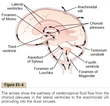
from the third ventricle, it flows downward along the aqueduct of Sylvius into the fourth ventricle, where still another minute amount of fluid is added. Finally, the fluid passes out of the fourth ventricle through three small openings, two lateral foramina of Luschkaand a midline foramen of Magendie, entering the cisterna magna, a fluid space that lies behind the medulla and beneath the cerebellum.
The cisterna magna is continuous with the subarach- noid space that surrounds the entire brain and spinal cord. Almost all the cerebrospinal fluid then flows upward from the cisterna magna through the subarach- noid spaces surrounding the cerebrum. From here, the fluid flows into and through multiple arachnoidal villi that project into the large sagittal venous sinus and other venous sinuses of the cerebrum. Thus, any extra fluid empties into the venous blood through pores of these villi.
Secretion by the Choroid Plexus. Thechoroid plexus, asection of which is shown in Figure 61–5, is a cauliflower-like growth of blood vessels covered by a thin layer of epithelial cells. This plexus projects into (1 and 2) the temporal horn of each lateral ventricle, (3) the posterior portion of the third ventricle, and (4) the roof of the fourth ventricle.
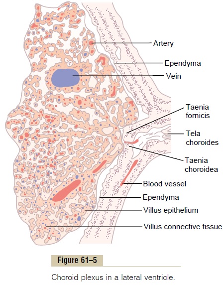
Secretion of fluid into the ventricles by the choroid plexus depends mainly on active transport of sodium ions through the epithelial cells lining the outside of the plexus.The sodium ions in turn pull along large amounts of chloride ions as well because the positive charge of the sodium ion attracts the chloride ion’s negative charge. The two of these together increase the quantity of osmotically active sodium chloride in the cere-brospinal fluid, which then causes almost immediate osmosis of water through the membrane, thus providing the fluid of the secretion.
Less important transport processes move small amounts of glucose into the cerebrospinal fluid and both potassium and bicarbonate ions out of the cerebrospinal fluid into the capillaries. Therefore, the resulting char-acteristics of the cerebrospinal fluid become the follow-ing: osmotic pressure, approximately equal to that of plasma; sodium ion concentration, also approximately equal to that of plasma; chloride ion, about 15 per cent greater than in plasma; potassium ion, approximately 40 per cent less; and glucose, about 30 per cent less.
Absorption of Cerebrospinal Fluid Through the Arachnoidal Villi.
The arachnoidal villi are microscopic fingerlike inward projections of the arachnoidal membrane through the walls and into the venous sinuses. Conglomerates of these villi form macroscopic structures called arach-noidal granulations that can be seen protruding into thesinuses. The endothelial cells covering the villi have been shown by electron microscopy to have vesicular passages directly through the bodies of the cells large enough to allow relatively free flow of (1) cerebrospinal fluid, (2) dissolved protein molecules, and (3) even par-ticles as large as red and white blood cells into the venous blood.
Perivascular Spaces and Cerebrospinal Fluid. The large arter-ies and veins of the brain lie on the surface of the brain but their ends penetrate inward, carrying with them a layer of pia mater, the membrane that covers the brain, as shown in Figure 61–6. The pia is only loosely adher-ent to the vessels, so that a space, the perivascular space, exists between it and each vessel. Therefore, perivascu-lar spaces follow both the arteries and the veins into the brain as far as the arterioles and venules go.
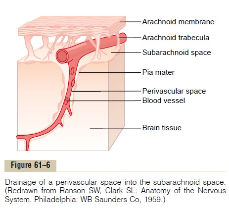
Lymphatic Function of the Perivascular Spaces. As is trueelsewhere in the body, a small amount of protein leaks out of the brain capillaries into the interstitial spaces of the brain. Because no true lymphatics are present in brain tissue, excess protein in the brain tissue leaves the tissue flowing with fluid through the perivascular spaces into the subarachnoid spaces. On reaching the sub-arachnoid spaces, the protein then flows with the cere-brospinal fluid, to be absorbed through the arachnoidalvilli into the large cerebral veins. Therefore, perivascu
In addition to transporting fluid and proteins, the perivascular spaces transport extraneous particulate matter out of the brain. For instance, whenever infec-tion occurs in the brain, dead white blood cells and other infectious debris are carried away through the perivascular spaces.
Related Topics