Chapter: Essentials of Anatomy and Physiology: Cell Structures and their Functions
Whole cell activity
Whole cell activity
A. Describe the process of gene expression.
B. Explain what is accomplished during mitosis.
C. Define differentiation, and explain how different celltypes develop.
A cell’s characteristics are ultimately determined by the types of proteins it produces. The proteins produced are in turn determined by the genetic information in the nucleus. In order to understand how a cell functions, we must consider the relationship between genes and proteins. For example, the transport of many food molecules into the cell requires cell membrane \proteins. Amino acids are assembled to synthesize proteins, including the transport proteins of the cell membrane. Information contained in DNA within the nucleus determines which amino acids are combined at ribosomes to form proteins. As you learned in the beginning, the human body is composed of trillions of cells, with many different characteristics\. Each human begins life as a single cell. Through cell division and cell differentiation, the cells that make up the human body are formed. The following sections illustrate the whole-cell activities that determine the characteris-tics of a functioning cell and the growth and maintenance of the human body.
Gene Expression
DNA contains the information that directs protein synthesis. This process is called gene expression. The proteins produced in a cell include those that serve as structural components inside the cell, proteins secreted to the outside of the cell, and enzymes that regulate chemical reactions in the cell. DNA influences the structural and functional characteristics of the entire organism because it directs protein synthesis. Whether an individual has blue eyes, brown hair, or other inherited traits is determined ultimately by DNA.
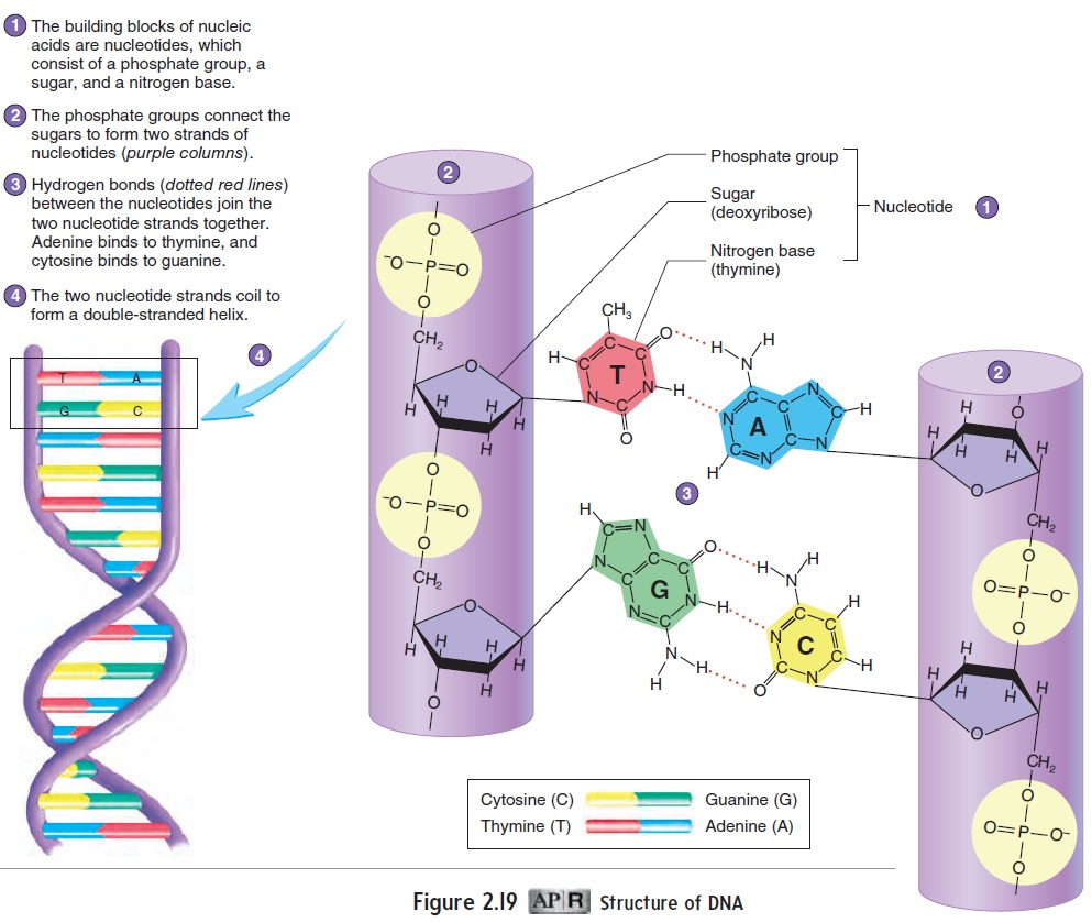
A DNA molecule consists of nucleotides joined together to form two nucleotide strands (see figures 2.19 and 3.23). The two strands are connected and resemble a ladder that is twisted aroundits long axis. The nucleotides function as chemical “letters” that form chemical “words.” A gene is a sequence of nucleotides (making a word) that provides a chemical set of instructions for making a specific protein. Each DNA molecule contains many different genes.

Recall that proteins consist of amino acids. The unique structural and functional characteristics of different proteins are determined by the kinds, numbers, and arrangement of their amino acids. The nucleotide sequence of a gene deter-mines the amino acid sequence of a specific protein.
Gene expression involves two steps—transcription and trans-lation (figure 3.22). This process can be illustrated with an analogy. Suppose a chef wants a cake recipe that is found only in a cookbook in the library. Because the book cannot be checked out, the chef makes a copy, or transcription, of the recipe. Later in the kitchen, the information contained in the copied recipe is used to make the cake. The changing of something from one form to another (from recipe to cake) is called translation.
In terms of this analogy, DNA (the cookbook) contains many genes (recipes) for making different proteins (food items). DNA, however, is too large a molecule to pass through the nuclear pores to the ribosomes (kitchen) where the proteins are made. Just as a book stays in the library, DNA remains in the nucleus. Therefore, through transcription, the cell makes a copy of the gene necessary to make a particular protein. The copy, which is called messengerRNA (mRNA), travels from the nucleus to the ribosomes in thecytoplasm, where the information in the copy is used to construct a protein by means of translation. Of course, the actual ingredients are needed to turn a recipe into a cake. The ingredients necessary to synthesize a protein are amino acids. Specialized molecules, called transfer RNAs (tRNAs),carry the amino acids to the ribo-some (figure 3.22).
In summary, gene expression involves transcription (making a copy of a gene) and translation (converting that copied informa-tion into a protein). Next, we consider the details of transcription and translation.
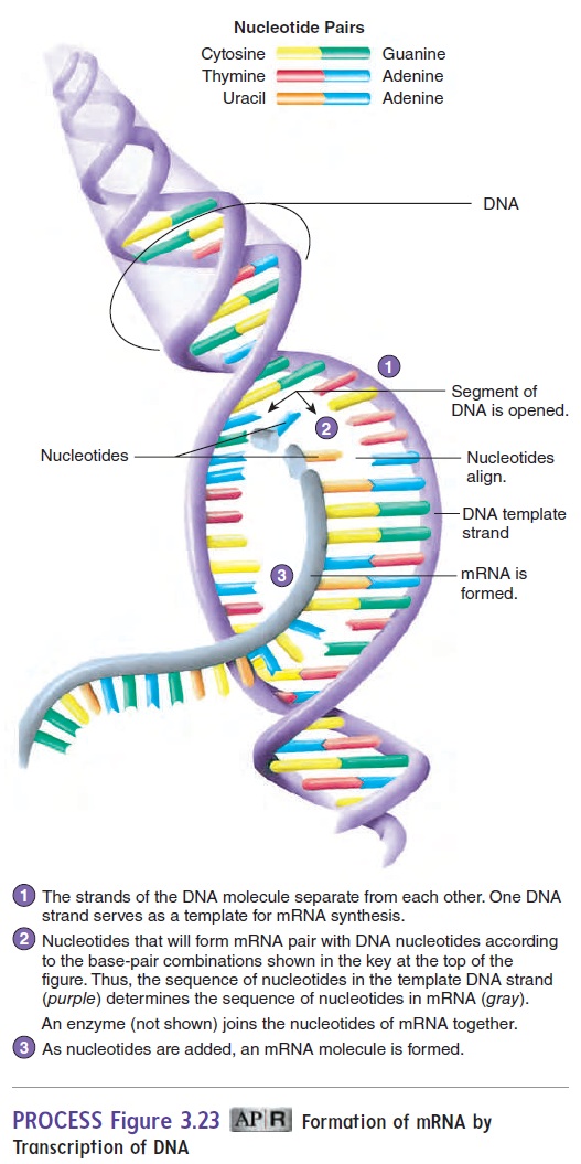
Transcription
The first step in gene expression, transcription, takes place in the nucleus of the cell (figure 3.22, steps 1 and 2). DNA determines the structure of mRNA through transcription. The double strands of a DNA segment separate, and DNA nucleotides pair with RNA nucleotides (figure 3.23, steps 1 and 2). Each nucleotide of DNA contains one of the following organic bases: thymine, adenine, cytosine, or guanine; each nucleotide of mRNA contains uracil, adenine, cytosine, or guanine. The number and sequence of nucleotides in the DNA serve as a template to determine the number and sequence of nucleotides in the mRNA. DNA nucleo-tides pair only with specific RNA nucleotides: DNA’s thymine with RNA’s adenine, DNA’s adenine with RNA’s uracil, DNA’s cytosine with RNA’s guanine, and DNA’s guanine with RNA’s cytosine.
After the DNA nucleotides pair up with the RNA nucleotides, an enzyme catalyzes reactions that form chemical bonds between the RNA nucleotides to form a long mRNA segment (figure 3.23, step 3). Once the mRNA segment has been transcribed, portions of the mRNA molecule may be removed.
The information in mRNA is carried in groups of three nucleotides called codons, which specify a particular amino acid. For example, the nucleotide sequence uracil, cytosine, and adenine (UCA) specifies the amino acid serine. There are 64 possible mRNA codons, but only 20 amino acids. As a result, more than 1 codon can specify the same amino acid. For example, CGA, CGG, CGU, and CGC code for the amino acid arginine; UUU and UUC code for phenylalanine. Some codons do not specify a particular amino acid but perform other functions. For example, UAA does not code for an amino acid. It is called a stop codon because it acts as a signal to end the translation process.
Translation
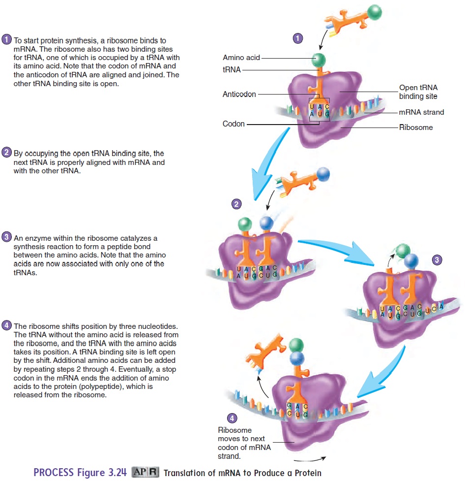
Translation, the synthesis of proteins based on the information in mRNA, occurs at ribosomes (see figure 3.22, steps 3–5). The mRNA molecules produced by transcription pass through the nuclear pores to the ribosomes. Ribosomes consist of small and large subunits, which combine with mRNA during translation. The process of translation requires two types of RNA in addition to the mRNA: tRNA andribosomal RNA (rRNA). There is one type of tRNA for each mRNA codon. The anticodon, a series of three nucleotides of tRNA, pairs with the codon of the mRNA. An amino acid is bound to another part of the tRNA. This ensures that the correct amino acid is matched with the codon of the mRNA. For example, the tRNA that pairs with the UUU codon of mRNA has the anticodon AAA and has the amino acid phenyl-alanine bound to it.
During translation, a ribosome binds to an mRNA. The ribo-some aligns the mRNA with tRNA molecules so that the anti-codons of tRNA can pair with the appropriate codons on the mRNA (figure 3.24, steps 1 and 2). An enzyme associated with the ribosome causes the formation of a peptide bond between the amino acids bound to the tRNAs (figure 3.24, step 3). The ribosome moves down the mRNA one codon at a time, releasing one of the tRNA and allowing the next tRNA to move into posi-tion. As the process continues, a polypeptide chain is formed. Translation ends when the ribosome reaches the stop codon on the mRNA (figure 3.24, step 4). The polypeptide chain is released and becomes folded to form the three-dimensional structure of the protein molecule (see figure 2.16). A protein can consist of a single polypeptide chain or two or more polypeptide chains that are joined after each chain is produced on a separate ribosome.
Cell Life Cycle
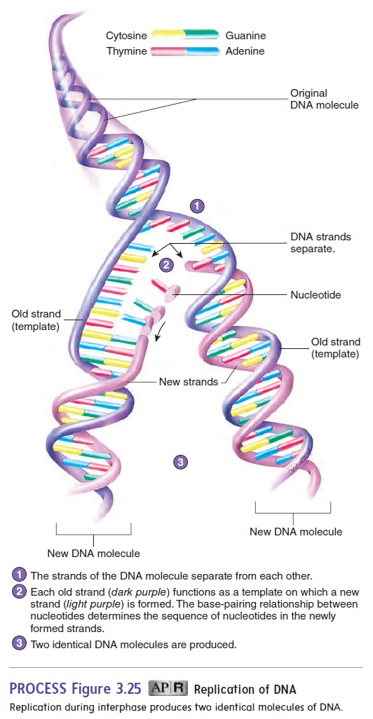
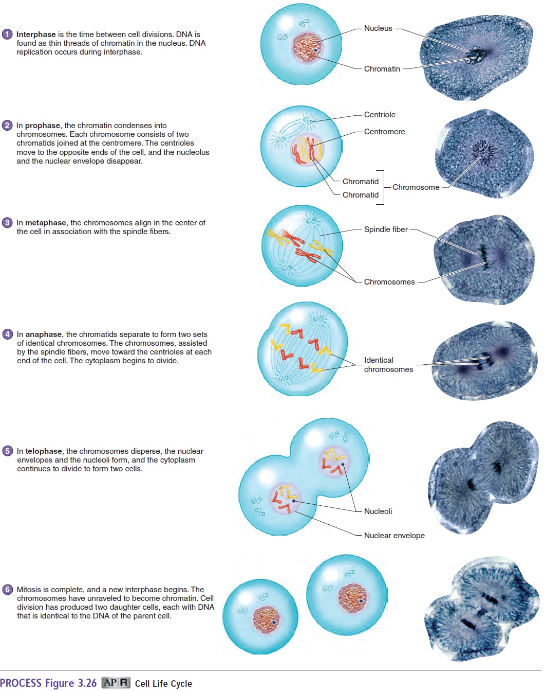
During growth and development, cell division allows for a dramatic increase in cell number after fertilization of an oocyte. The same process allows for the replacement and repair of damaged tissue. The cell life cycle includes two major phases: a nondividing phase, called interphase, and cell division. A cell spends most of its life cycle in interphase performing its normal functions. During interphase, the DNA (located in chromosomes in the cell’s nucleus) is replicated. The two strands of DNA separate from each other, and each strand serves as a template for the production of a new strand of DNA (figure 3.25). Nucleotides in the DNA of each template strand pair with nucleotides that are subsequently joined by enzymes to form a new strand of DNA. The sequence of nucleotides in the DNA template determines the sequence of nucleotides in the new strand of DNA because adenine pairs with thymine, and cytosine pairs with guanine. The two new strands of DNA combine with the two template strands to form two double strands of DNA.
At the end of interphase, a cell has two complete sets of genetic material. The DNA is dispersed throughout the nucleus as thin threads called chromatin (figure 3.26, step 1; see also figures 3.13b and 3.14).
Cell division is the formation of daughter cells from a single parent cell. The new cells necessary for growth and tissue repair are formed through mitosis (discussed next), and the sex cells necessary for reproduction are formed through meiosis.
Each cell of the human body, except for sex cells, contains 46 chromosomes. Sex cells have half the number of chromosomesas other cells (see “Meiosis”). The 46 chromosomes are the diploid (dip′loyd) number of chromosomes and are organizedto form 23 pairs of chromosomes. Of the 23 pairs, 1 pair is the sex chromosomes, which consist of 2 X chromosomes if the person is a female or an X chromosome and a Y chromosome if the person is a male. The remaining 22 pairs of chromosomes are called autosomes\ (aw′tō-sōmz). The sex chromosomes determine the individual’s sex, and the autosomes determine most other characteristics.
Mitosis
Most cells of the body, except those that give rise to sex cells, divide by mitosis (mı̄-tō′sis). During mitosis, a parent cell divides to form two daughter cells with the same amount and type of DNA as the parent cell. Because DNA determines the structure and function of cells, the daughter cells, which have the same DNA as the parent cell, can have the same structure and perform the same functions as the parent cell.
For convenience, mitosis is divided into four stages: prophase, metaphase, anaphase, and telophase (figure 3.26, steps 2–5). Although each stage represents certain major events, the process of mitosis is continuous. Learning each of the stages is helpful, but the most important concept to understand is how each of the two cells produced by mitosis obtains the same number and type of chromosomes as the parent cell.
· Prophase. Duringprophase(figure 3.26, step 2), thechromatin condenses to form visible chromosomes. After
interphase, each chromosome is made up of two genetically identical strands of chromatin, called chromatids (krō′mă-tidz), which are linked at one point by a specialized region called the centromere(sen′trō-mēr; kentron, center + meros, part). Replication of the genetic material duringinterphase results in the two identical chromatids of each chromosome. Also during prophase, microtubules called spindle fibers extend from the centrioles to the centromeres (see figures 3.1 and 3.21). The centrioles divide and migrate to each pole of the cell. In late prophase, the nucleolus and nuclear envelope disappear.
· Metaphase. Inmetaphase(figure 3.26, step 3), thechromosomes align near the center of the cell.
· Anaphase. At the beginning ofanaphase(figure 3.26,step 4), the chromatids separate. When this happens, each chromatid is then called a chromosome. At this point, two identical sets of 46 chromosomes are present in the cell. Each of the two sets of 46 chromosomes is moved by the spindle fibers toward the centriole at one of the poles of the cell. At the end of anaphase, each set of chromosomes has reached an opposite pole of the cell, and the cytoplasm begins to divide.
· Telophase. Duringtelophase(tel′ō-fāz) (figure 3.26, step 5),the chromosomes in each of the daughter cells become organized to form two separate nuclei. The chromosomes begin to unravel and resemble the genetic material during interphase.
Following telophase, cytoplasm division is completed, and two separate daughter cells are produced (figure 3.26, step 6).
Differentiation
A sperm cell and an oocyte unite to form a single cell, and a new individual begins. The single cell formed during fertiliza-tion divides by mitosis to form two cells, which divide to form four cells, and so on . The trillions of cells that ultimately make up the body of an adult, as a result, stem from that single cell. Therefore, all the cells in an individual’s body contain the same amount and type of DNA. But even though the genetic information contained in cells is identical, not all cells look and function alike. Bone cells, for example, do not look like or function the same as muscle cells, nerve cells, or red blood cells (figure 3.27).
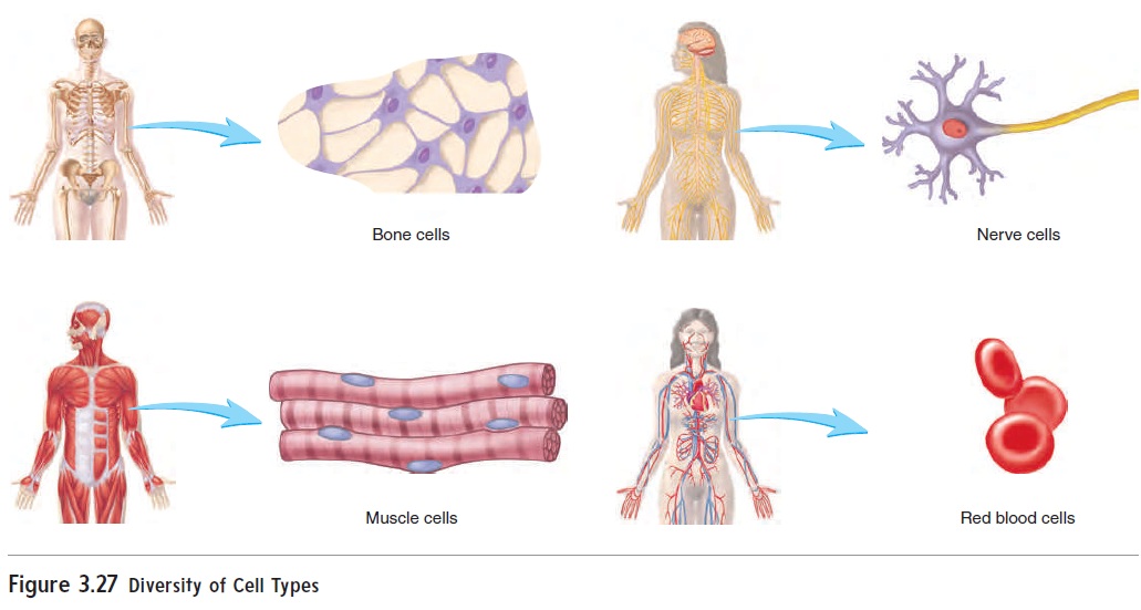
The process by which cells develop with specialized structures and functions is called differentiation. During differentiation of a cell, some portions of DNA are active, but others are inactive. The active and inactive sections of DNA differ with each cell type. For example, the portion of DNA responsible for the structure and function of a bone cell is different from that responsible for the structure and function of a muscle cell. Differentiation, then, results from the selective activation and inactivation of segments of DNA. The mechanisms that determine which portions of DNA are active in any one cell type are not fully understood, but the resulting differentiation produces the many cell types that function together to make a person. Eventually, as cells differentiate and mature, the rate at which they divide slows or even stops.
Apoptosis
Apoptosis (ăp′\op-tō′\sis), orprogrammed cell death,is a normalprocess by which cell numbers within various tissues are adjusted and controlled. In the developing fetus, apoptosis removes extra tissue, such as cells between the developing fingers and toes. In some adult tissues, apoptosis eliminates excess cells to maintain a constant number of cells within the tissue. Damaged or potentially dangerous cells, virus-infected cells, and potential cancer cells are also eliminated by apoptosis.
Apoptosis is regulated by specific genes. The proteins coded for by those genes initiate events within the cell that ultimately lead to the cell’s death. As apoptosis begins, the chromatin within the nucleus condenses and fragments. This is followed by frag-mentation of the nucleus and finally by death and fragmentation of the cell. Specialized cells called macrophages phagocytize the cell fragments.
Related Topics