Chapter: Biotechnology Applying the Genetic Revolution: Immune Technology
Visualizing Cell Components Using Antibodies
VISUALIZING
CELL COMPONENTS USING ANTIBODIES
Antibodies can be used to
visualize the location of specific proteins within the cell. Immunocytochemistry refers to the
visualization of specific antigens in cultured cells, whereas immunohistochemistry
refers to their visualization in prepared tissue sections. In either technique,
the first step is to prepare the cells. They must be treated to maintain their
cellular architecture, so that the cells appear much as they would if still
alive. Usually, the cells are treated with crosslinking agents such as
formaldehyde or with denaturants like acetone or methanol. In
immunohistochemistry, tissue samples can be frozen and then sliced into small
thin sections (about 4 mm), providing a two-dimensional view of the cell.
Another option is to embed the tissue sample in paraffin wax. Here the cells
are first dehydrated in a series of alcohol solutions, and then treated with
the wax. The tissue is then sectioned into thin two-dimensional slices as for
frozen tissues.
Preserved cells then need to
be permeabilized so that the antibody can enter. Once a single, thin layer of
prepared cells or tissue sections is readied, the cells are treated to make the
antigen more accessible to the antibody. If in wax, the tissue sections are
dewaxed and rehydrated in solution. Fixed tissue sections can be irradiated
with microwaves, which break the crosslinks induced by the fixative, or the
samples can be heated under pressure. After permeabilization, the primary
antibody finds its antigen within the sample and binds. A secondary antibody
contains the detection system. The secondary antibody binds to the primary
antibody/antigen complex, and then the appropriate reagents are added to
visualize the location of the complex. (In some cases, a single antibody, with
an attached detection system, is used.)
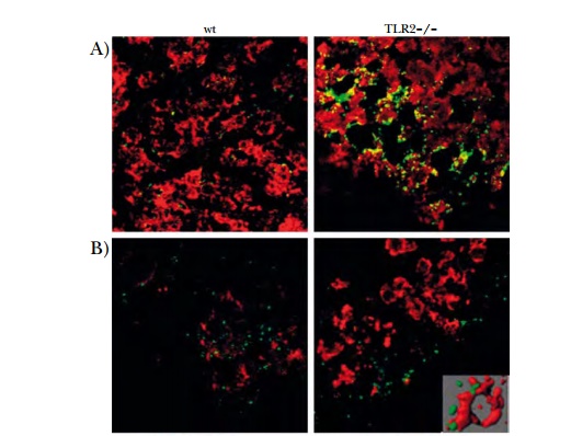
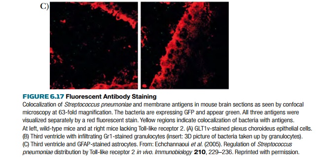
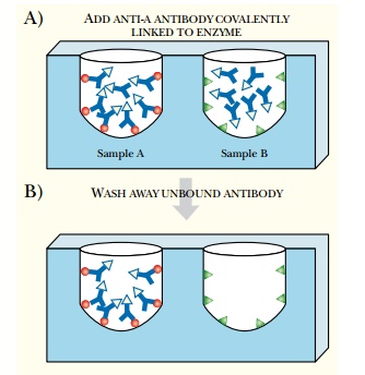
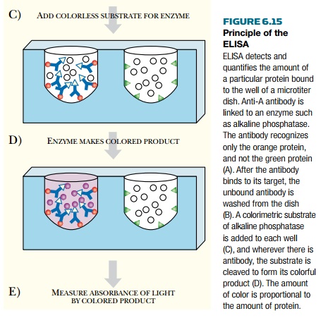
The antibody is detected using enzymes or by fluorescent labels. A common enzyme-mediated detection system is alkaline phosphatase, as with the ELISA—see Fig. 6.15. Fluorescently labeled antibodies must be excited with UV light, on which the fluorescent label emits light at a longer wavelength. Samples are directly visualized with a microscope attached to a UV light source (Fig. 6.17). Fluorescent antibodies tend to bleach out when exposed to excess UV; therefore, the microscope is attached to a camera to record the data as a digital image.
Related Topics