Chapter: Biotechnology Applying the Genetic Revolution: Nanobiotechnology
Visualization at the Nanoscale
VISUALIZATION
AT THE NANOSCALE
In order to manipulate matter
on an atomic scale, we need to see individual atoms and molecules. Although
individual molecules have been visualized with the electron microscope, it was
the development of scanning probe microscopes that opened up the field of
nanotechnology. These instruments all rely on a miniature probe that scans
across the surface under investigation.
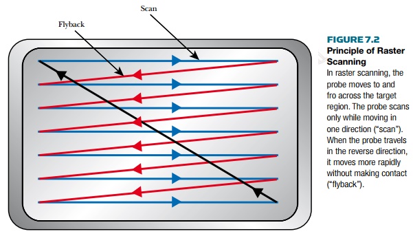
All scanning probe
microscopes work by measuring some property, such as electrical resistance,
magnetism, temperature, or light absorption, with a tip positioned extremely
close to the sample. The microscope raster-scans the probe over the sample
(Fig. 7.2) while measuring the property of interest.
The data are displayed as a
raster image similar to that on a television screen.
Unlike traditional
microscopes, scanned-probe systems do not use lenses, so the size of the probe
rather than diffraction limits their resolution. Some of these instruments can
be used to alter samples as well as visualize them.
The first of these
instruments was the scanning tunneling microscope (STM), which was developed by
Gerd Binnig and Heinrich Rohrer at IBM (see following section). They received
the Nobel Prize in 1986. The STM sends electrons, that is, an electric current,
through the sample and so measures electrical resistance. The atomic force
microscope (AFM) is especially useful in biology and measures the force between
the probe tip and the sample.
Related Topics