Chapter: Medical Physiology: The Eye: I. Optics of Vision
Visual Acuity - Optics of the Eye
Visual Acuity
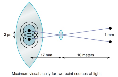
Theoretically, light from a distant point source, when focused on the retina, should be infinitely small. However, because the lens system of the eye is never perfect, such a retinal spot ordinarily has a total diam-eter of about 11 micrometers, even with maximal res-olution of the normal eye optical system. The spot is brightest in its center and shades off gradually toward the edges, as shown by the two-point images in Figure 49–16.
The average diameter of the cones in the fovea of the retina—the central part of the retina, where vision is most highly developed—is about 1.5 micrometers, which is one seventh the diameter of the spot of light.
Nevertheless, because the spot of light has a bright center point and shaded edges, a person can normally distinguish two separate points if their centers lie as much as 2 micrometers apart on the retina, which is slightly greater than the width of a foveal cone. This discrimination between points is also shown in Figure 49–16.
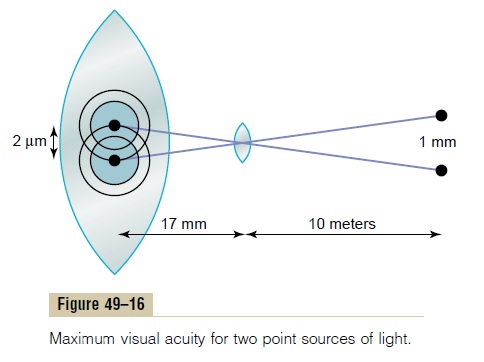
The normal visual acuity of the human eye for dis-criminating between point sources of light is about 25 seconds of arc. That is, when light rays from two sepa-rate points strike the eye with an angle of at least 25 seconds between them, they can usually be recognized as two points instead of one. This means that a person with normal visual acuity looking at two bright pin-point spots of light 10 meters away can barely distin-guish the spots as separate entities when they are 1.5 to 2 millimeters apart.
The fovea is less than 0.5 millimeter (less than 500 micrometers) in diameter, which means that maximum visual acuity occurs in less than 2 degrees of the visual field. Outside this foveal area, the visual acuity becomes progressively poorer, decreasing more than 10-fold as the periphery is approached. This is caused by the connection of more and more rods and cones to each optic nerve fiber in the nonfoveal, more peripheral parts of the retina.
Clinical Method for Stating Visual Acuity. The chart fortesting eyes usually consists of letters of different sizes placed 20 feet away from the person being tested. If the person can see well the letters of a size that he or she should be able to see at 20 feet, the person is said to have 20/20 vision—that is, normal vision. If the person can see only letters that he or she should be able to see at 200 feet, the person is said to have 20/200 vision. In other words, the clinical method for express-ing visual acuity is to use a mathematical fraction that expresses the ratio of two distances, which is also the ratio of one’s visual acuity to that of a person with normal visual acuity.
Related Topics