Chapter: Essentials of Anatomy and Physiology: Muscular System
Upper limb Muscles
Upper limb Muscles
The muscles of the upper limb include those that attach the limb and pectoral girdle to the body and those in the arm, forearm, and hand.
Scapular Movements
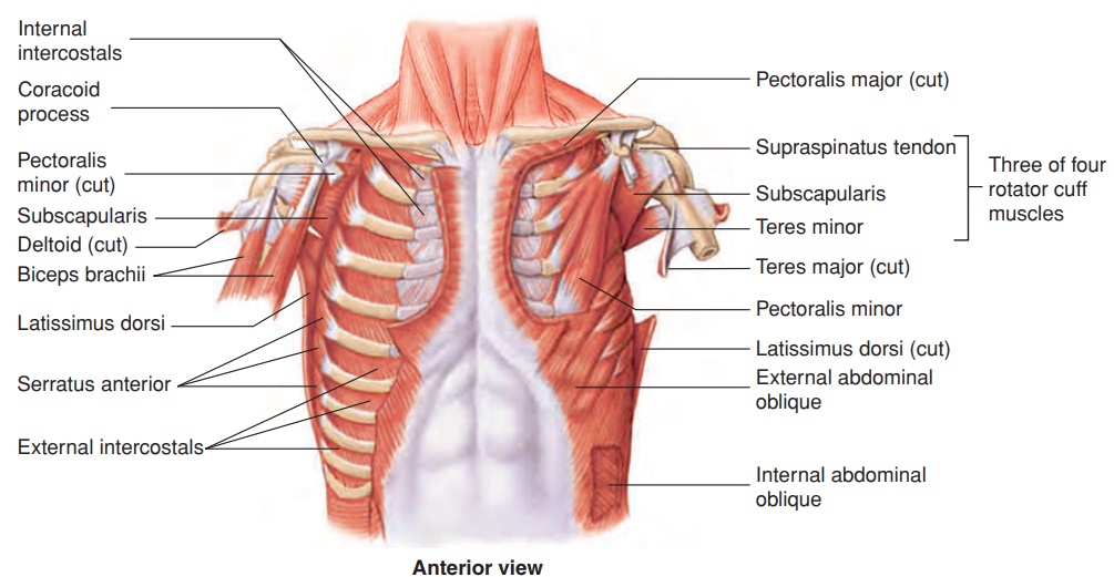
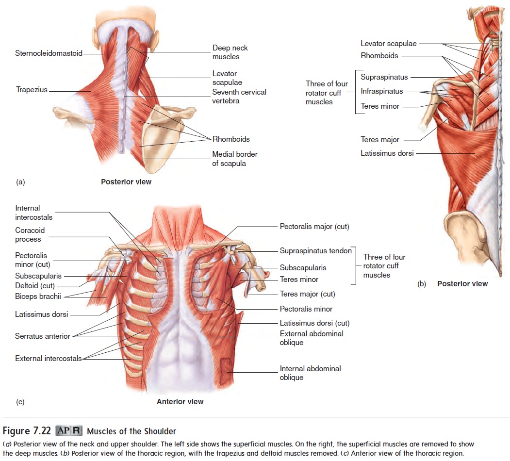
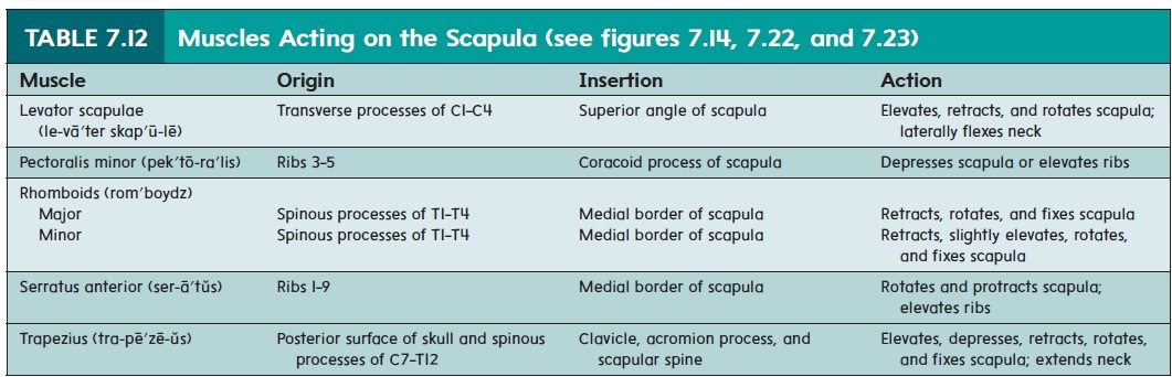
The upper limb is primarily connected to the body by muscles. The muscles that attach the scapula to the thorax and move the scapula include the trapezius (tra-pē′zē-ŭs), the levator scapulae (le-vā′ter skap′ū-lē), the rhomboids (rom′boydz), the serratus (ser-ā′tŭs; serrated) anterior, and the pectoralis (pek′tō-ra′lis) minor (figure 7.22 and table 7.12). These muscles act as fixatorsto hold the scapula firmly in position when the muscles of the arm contract. The scapular muscles also move the scapula into differentpositions, thereby increasing the range of movement of the upper limb. The trapezius forms the upper line from each shoulder to the neck. The origin of the serratus anterior from the first eight or nine ribs can be seen along the lateral thorax.
Arm Movements
The arm is attached to the thorax by the pectoralis major and latissimus dorsi (lă-tis′i-mŭs dōr′sı̄) muscles (figure 7.23aand table 7.13; see figure 7.22c). The pectoralis major adducts the arm and flexes the shoulder. It can also extend the shoulder from a flexed position. The latissimus dorsi medially rotates and adducts the arm and powerfully extends the shoulder. Because a swimmer uses these three motions during the power stroke of the crawl, the latissimus dorsi is often called the swimmer’s muscle.
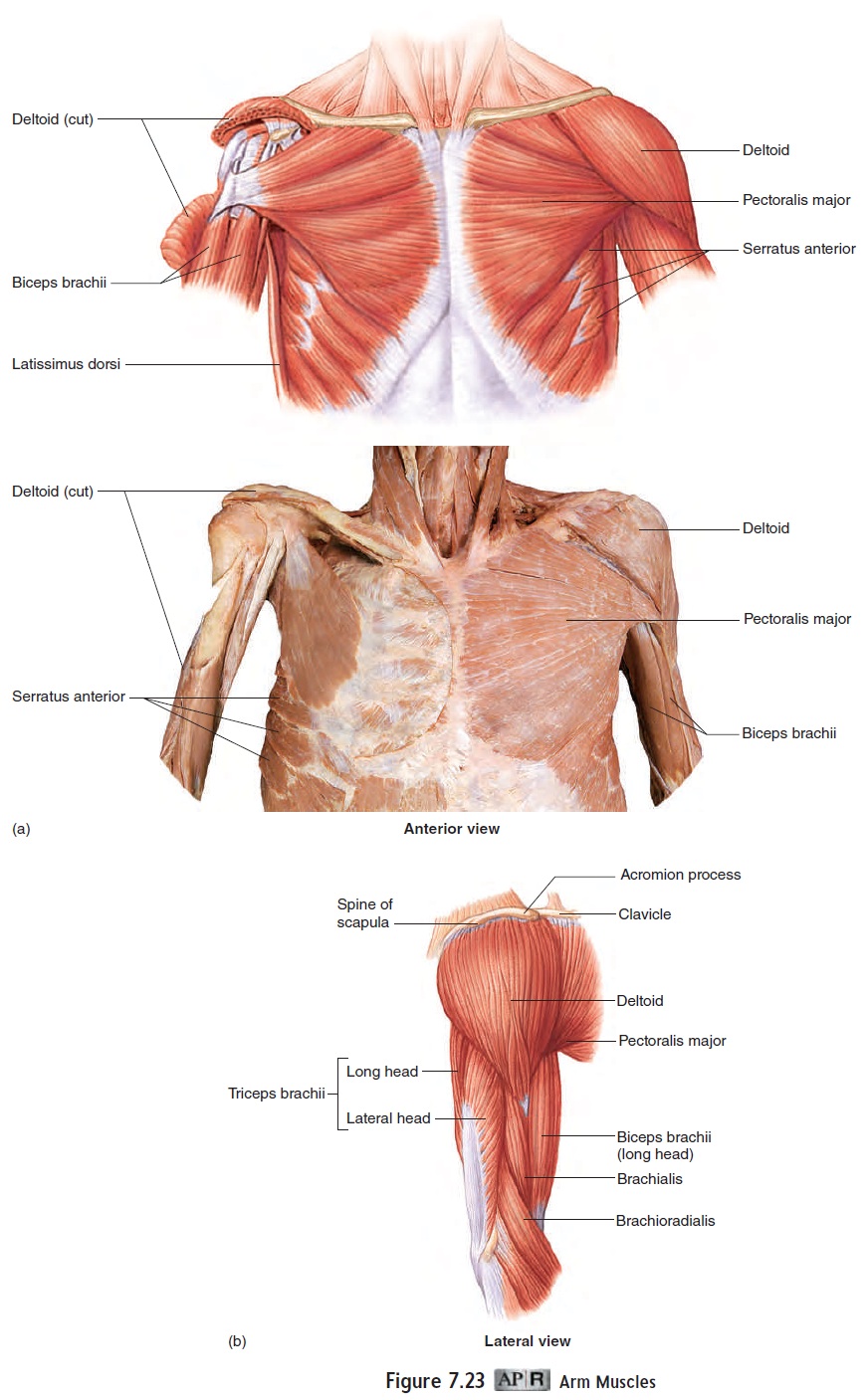
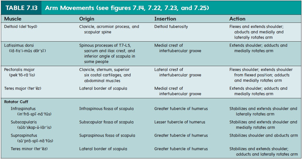
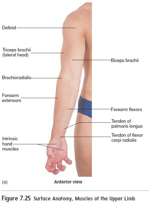
Another group of four muscles, called the rotator cuff muscles, attaches the humerus to the scapula and forms a cuff or cap over the proximal humerus (table 7.13; see figure 7.22b,c). These muscles stabilize the joint by holding the head of the humerus in the glenoid cavity during arm movements, especially abduction. A rotator cuff injury involves damage to one or more of these muscles or their tendons. Thedeltoid (del′toyd) muscle attaches the humerus to the scapula and clavicle and is the major abductor of the upper limb. The pectoralis major forms the upper chest, and the deltoid forms the rounded mass of the shoulder (see figure 7.25). The deltoid is a common site for administering injections.
Forearm Movements
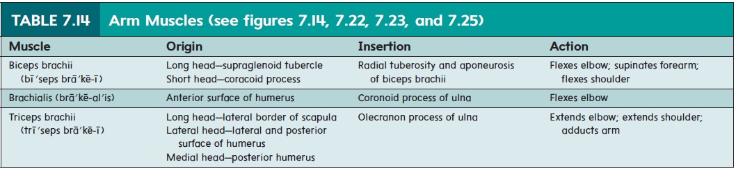
The arm can be divided into anterior and posterior compartments. The triceps brachii (trı̄′seps brā′kē-ı̄; three heads, arm), the pri-mary extensor of the elbow, occupies the posterior compartment (figure 7.23band table 7.14). The anterior compartment is occupied mostly by the biceps (bı̄′seps) brachii and the brachialis (brā′kē-ăl-is), the primary flexors of the elbow. The brachioradialis (brā′kē-ō-rā′dē-al′is), which is actually a posterior forearm muscle, helps flex the elbow.
Supination and Pronation
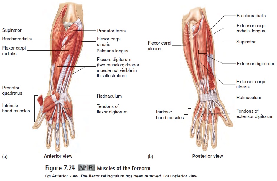
Supination of the forearm, or turning the flexed forearm so that the palm is up, is accomplished by the supinator (soo′pi-nā-ter) (figure 7.24 and table 7.15) and the biceps brachii, which tends to supinate the forearm while flexing the elbow. Pronation, turning the forearm so that the palm is down, is a function of two prona-tor (prō-nā′ter) muscles.
Wrist and Finger Movements
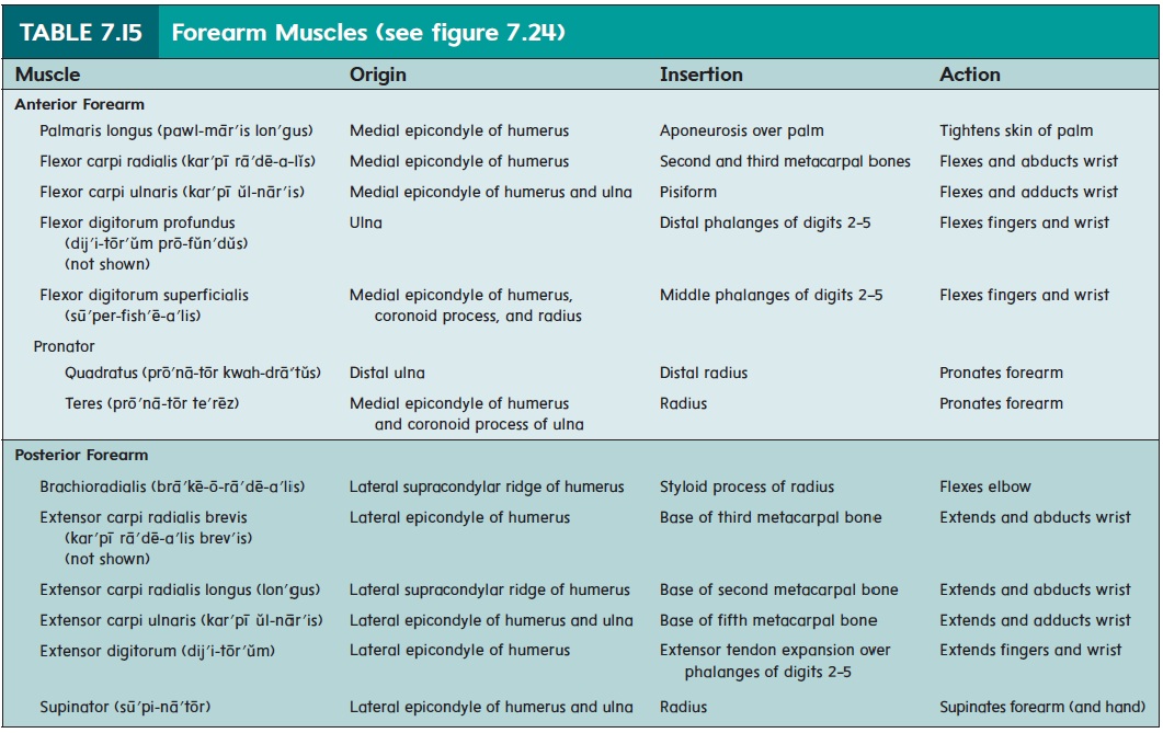
The twenty muscles of the forearm can also be divided into anterior and posterior groups. Only a few of these muscles, the most superficial, appear in table 7.15 and figure 7.24. Most of the anterior forearm muscles are responsible for flexion of the wrist and fingers, whereas most of the posterior forearm muscles cause extension.
A strong band of fibrous connective tissue, the retinaculum (figure 7.24b), covers the flexor and extensor ten-dons and holds them in place around the wrist so that they do not “bowstring” during muscle contraction. Because the retinaculum does not stretch as a result of pressure, this characteristic is a contributing factor in carpal tunnel syndrome
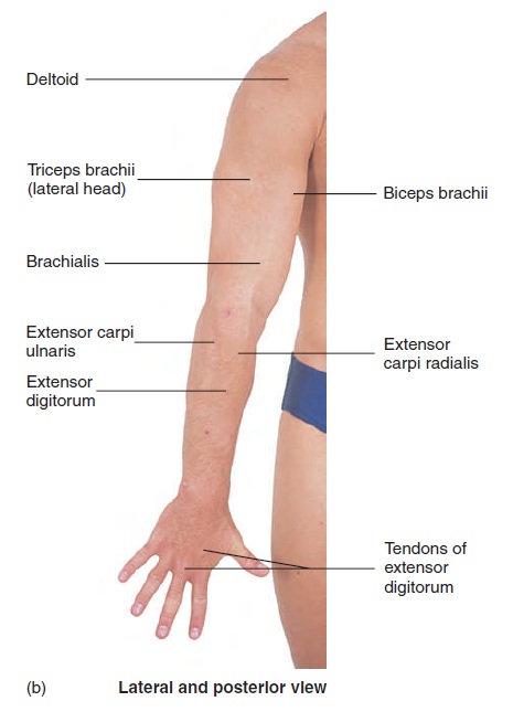
The flexor carpi (kar′ pı̄) muscles flex the wrist, and the extensor carpi muscles extend the wrist. The tendon of the flexorcarpi radialis serves as a landmark for locating the radial pulse (figure 7.25a). The tendons of the wrist extensors are visible on the posterior surface of the forearm (figure 7.25b). Forceful, repeated contraction of the wrist extensor muscles, as occurs in a tennis backhand, may result in inflammation and pain where the extensor muscles attach to the lateral humeral epicondyle. This condition is sometimes referred to as “tennis elbow.” Flexion of the fingers is the function of the flexor digitorum (dij′ i-tōr′ ŭm;flexor of the digits, or fingers). Extension of the fingers is accom-plished by the extensor digitorum. The tendons of this muscle are very visible on the dorsal surface of the hand (figure 7.25b). The thumb has its own set of flexors, extensors, adductors, and abduc-tors. The little finger has some similar muscles.
Nineteen muscles, called intrinsic hand muscles, are locat-ed within the hand. Interossei (in′ ter-os′ ē-ı̄; between bones) muscles, located between the metacarpal bones, are responsible for abduction and adduction of the fingers. Other intrinsic hand muscles are involved in the many possible movements of the thumb and fingers. These muscles account for the fleshy masses at the base of the thumb and little finger and the fleshy region between the metacarpal bones of the thumb and index finger.
Related Topics