Chapter: Human Neuroanatomy(Fundamental and Clinical): The Olfactory and Limbic Regions
The Olfactory Region
The Olfactory Region
A major difficulty in considering the olfactory pathways is that these involve numerous, rather obscure and small, structures and areas. For a proper understanding it is necessary that the names and positions of these areas be introduced at the outset.
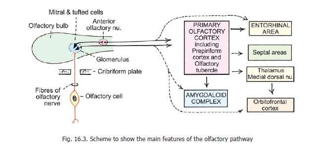
The peripheral end organ for smell is the olfactory mucosa that lines the upper and posterior parts of the nasal cavity. Nerve fibres arising in this mucosa collect to form about twenty bundles that together constitute an olfactory nerve. The bundles pass through foramina in the cribriform plate of the ethmoid bone to enter the cranial cavity where they terminate in the olfactory bulb (Figs. 16.1, 16.3).
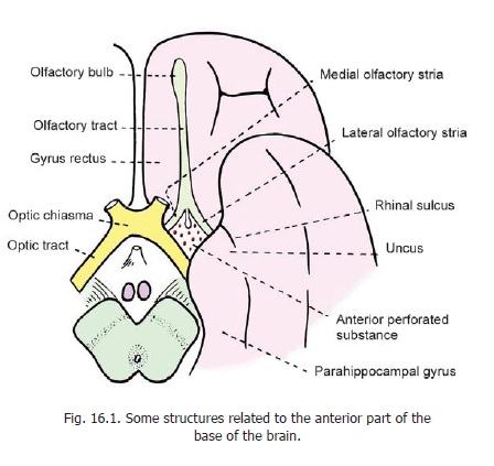
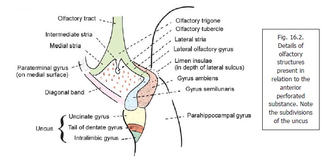
The olfactory bulb is an elongated oval structure that lies just above the cribriform plate. It is continuous posteriorly with the olfactory tract through which it is connected to the base of the cerebral hemisphere. When traced posteriorly the olfactory tract divides intomedial and lateralolfactory striae (Figs. 16.1, 16.2). The point of bifurcation is expanded and forms the olfactory trigone. An intermediate stria is sometimes present. The olfactory striae are intimately related toa mass of grey matter called the anterior perforated substance. The medial and lateral striae form the anteromedial and anterolateral boundaries of this substance. The intermediate stria extends into the anterior perforated substance and ends in a slight elevation (in the anterior part of the substance) called the olfactory tubercle. Posterolaterally, the anterior perforated substance is related to the uncus (Fig. 16.1), while posteromedially it is bounded by a bundle of fibres called the diagonalband (of Broca)(Fig. 16.2).
The uncus is a part of the cerebral hemisphere that lies on the tentorial surface a little behind and medial to the temporal pole. It represents the anterior end of theparahippocampal gyrus and is separated from the temporal pole by the rhinal sulcus.
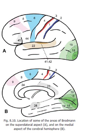
The anterior part of the parahippocampal gyrus, including the uncus, is referred to as the entorhinal area (area 28). Some other areas present in the region are shown in Fig. 16.2.
Advanced:
The uncus is subdivided into three parts. From anterior to posterior side these are (Fig. 16.2) the uncinate gyrus, the tail of the dentate gyrus (band of Giacomini) and theintralimbic gyrus.
When traced backwards the lateral olfactory stria reaches the limen insulae (in the depth of the stem of the lateral sulcus). Here it bends sharply to the medial side and becomes continuous with a small area of grey matter lying anterior to the uncus and called the gyrus semilunaris (or periamygdaloid area). The gyrus semilunaris is closely related to theamygdaloid complex which lies deep (i.e., superior) to it. The lateral olfactory stria is covered by a thin layer of grey matter called the lateral olfactory gyrus. When traced backwards this gyrus becomes continuous with a part of the cortex called the gyrus ambiens. The gyrus ambiens lies lateral to the gyrus semilunaris. Posteriorly, it becomes continuous with the entorhinal area. The lateral olfactory gyrus and the gyrus ambiens collectively form the prepiriform region (or area).
The term piriform lobe is applied collectively to the following parts that have been identified above.
1. Prepiriform region (lateral olfactory gyrus and gyrus ambiens).
2. Gyrus semilunaris (or periamygdaloid region).
3. Lateral olfactory stria (?).
4. Anterior part of parahippocampal gyrus, including the uncus (entorhinal area, Brodmann’s area 28: See Fig. 8.10).
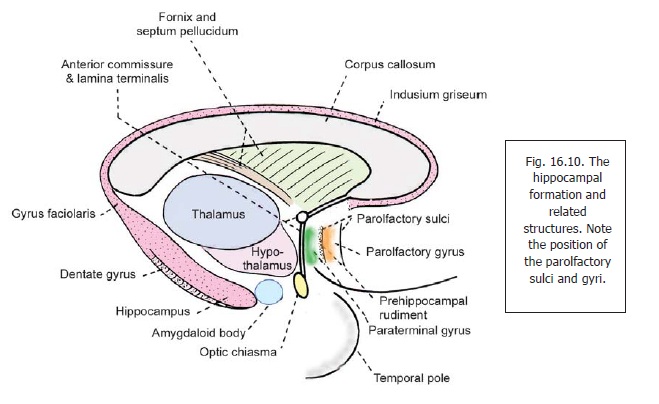
When traced medially, the medial olfactory stria, and the diagonal band, both reach the medial surface of the hemisphere, where they end near the paraterminal gyrus (which lies just in front of the lamina terminalis: Fig. 16.10).
Related Topics