Chapter: Human Neuroanatomy(Fundamental and Clinical): Ventricles of the Brain
The Lateral Ventricles - Ventricles of the Brain
The Lateral Ventricles
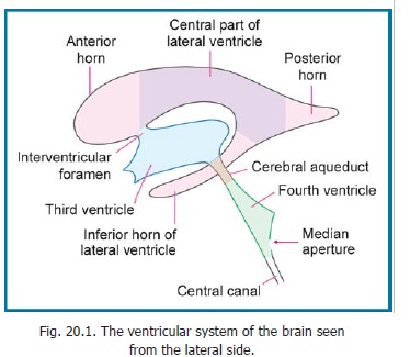
The lateral ventricles are two cavities, one situated within each cerebral hemisphere. Each ventricle consists of a central part which gives off three extensions called the anterior, posterior and inferior horns (Fig. 20.1).
The Central Part
The central part of the lateral ventricle is elongated anteroposteriorly. Anteriorly, it becomes continuous with the anterior horn, at the level of the interventricular foramen. Posteriorly, the central part reaches the splenium of the corpus callosum.
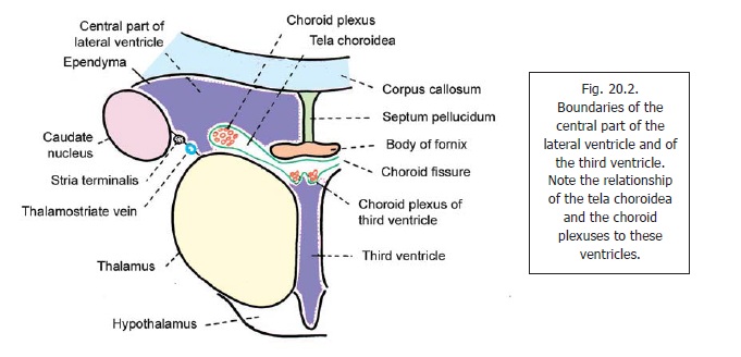
The central part is triangular in cross section (Fig. 20.2). It has a roof, a floor, and a medial wall. The roof and floor meet on the lateral side. The roof is formed by the trunk of the corpus callosum. The medial wall is formed by the septum pellucidum and by the body of the fornix. It is common to thetwo lateral ventricles. The floor is formed mainly by the superior surface of the thalamus (medially), and by the caudate nucleus (laterally). Between these two structures there are the stria terminalis(laterally) and the thalamostriate vein (medially). From Fig. 20.2 it will be seen that there is a space between the fornix and the upper surface of the thalamus. This is the choroid fissure.
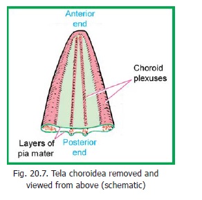
A fold of pia mater, the tela choroidea, invaginates into the ventricle through the fissure and covers part of the thalamus. The tela choroidea is common to the two lateral ventricles, and to the third ventricle. Within each lateral edge of the tela choroidea there are plexuses of blood vessels that constitute the choroid plexus (Figs. 20.2, 20.7). The tela choroidea and other structures forming the walls of theventricle are lined by ependyma.
The Anterior Horn
The anterior horn of the lateral ventricle, lies anterior to its central part, the two being separated by an imaginary vertical line drawn at the level of the interventricular foramen (Fig. 20.1). This horn is triangular in section. It has a roof, a floor and a medial wall (Fig. 20.3). It is closed, anteriorly, by the genu and rostrum of the corpus callosum.
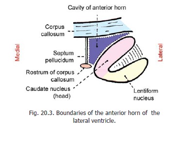
The roof is formed by the most anterior part of the trunk of the corpus callosum. The floor is formed mainly by the head of the caudate nucleus. A small part of the floor, near the middle line, is formed by the upper surface of the rostrum of the corpus callosum. The medial wall(common to the two sides) is formed by the septum pellucidum. It may be noted that the tela choroidea and the choroid plexus do not extend into the anterior horn.
The Posterior Horn
The posterior horn of the lateral ventricle extends backwards into the occipital lobe. It has a roof, a lateral wall, and a medial wall (Fig. 20.4).
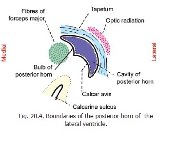
The roof and lateral wall are formed by the tapetum. The medial wall shows two elevations. The upper of these is the bulb of the posterior horn, which is produced by fibres of the forceps major as they run backwards from the splenium of the corpus callosum. The lower elevation is called the calcar avis. It represents white matter ‘pushed in’ by formation of the calcarine sulcus.
The Inferior Horn
The inferior horn of the lateral ventricle begins at the posterior end of the central part. It runs downwards and forwards into the temporal lobe, its anterior end reaching close to the uncus.
In considering the structures to be seen in the walls of the inferior horn it is useful to note that the anterior horn, the central part, and the inferior horn form one continuous C-shaped cavity. From Fig. 20.1 it will be obvious that the floor of the central part of the ventricle is continuous with the roof of the inferior horn. It is also useful to recall that the body of the fornix divides, posteriorly, into two crura which become continuous with the fimbria and hippocampus.
In the central part of the ventricle, the choroid fissure lies below the fornix. When traced into the inferior horn, the fissure lies above the fimbria and hippocampus. The choroid plexus extends into the inferior horn through the choroid fissure.
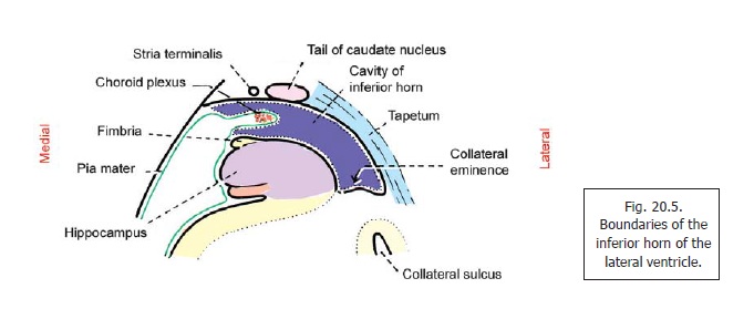
In cross section, the inferior horn is seen to have a narrow cavity (Fig. 20.5). The cavity is bounded above, and laterally, by the roof; and below, and medially, by the floor. (Because of this orientation the lateral part of the roof is sometimes called the lateral wall, and the medial part of the floor is called the medial wall.)
The lateral part of the roof (or lateral wall) is formed by fibres of the tapetum. The medial part of the roof is formed by the tail of the caudate nucleus (laterally) and the stria terminalis (medially). These structures are continued into the roof of the inferior horn from the floor of the central part. Anteriorly, the tail of the caudate nucleus and the stria terminalis end in relation to the amygdaloid complex, which lies in the most anterior part of the roof.
The floor of the inferior horn is formed mainly by the hippocampus, along with the alveus and fimbria. In the lateral part of the floor there is an elevation, the collateral eminence, produced by inward bulging of the white matter which lies deep to the collateral sulcus.
Related Topics