Chapter: 10th Science : Chapter 13 : Structural Organisation of Animals
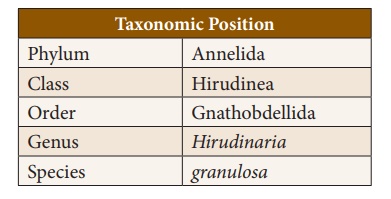
The Indian Cattle Leech (Hirudinaria granulosa)
The
Indian Cattle Leech (Hirudinaria
granulosa)
1. Habit and Habitat
Hirudinaria granulosa (Indian Cattle Leech)
is found in India, Bangladesh, Pakistan, Myanmar and Srilanka. It lives in
freshwater ponds, lakes, swamps and slow streams. They are ectoparasitic
and feed on the blood of fishes, frogs, cattle and human. It is sanguivorous
(blood sucking) in nature.
2. External Morphology
Shape and Size: The body of a leech is
soft, vermiform, elongated and segmented. It becomes ribbon shaped when
extended and almost cylindrical when contracted. Leeches may grow to a length
of 35cm.
Colouration: Dorsal surface is olive
green in colour and the ventral surface is orange yellow or orange red
in colour.
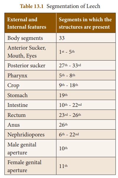
Segmentation: Metamerism
is the segmentation
of the body. The body of leech is metamerically divided into 33 segments.
The segments are arranged one behind the other. Each segment is further
superficially subdivided into rings or annuli. A temporary clitellum
is formed on segments 9-11, which is meant to produce a cocoon during
the breeding season.
Receptors: On the dorsal side there
are five pairs of eyes on the first five segments. Each segment bears a
number of sensory projections called receptors. Annular receptors are
located in each annulus and segmental receptors are located on the first
annulus of each segment.
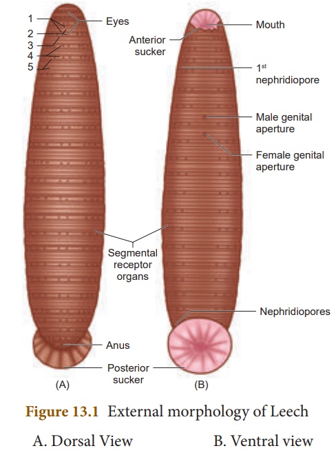
Suckers: Leech has two suckers.
The sucker located at the anterior end is called anterior sucker or
oral sucker which is ventral in position occupying the first five segments.
The posterior sucker is formed by the fusion of the last seven segments.
The anterior sucker helps in feeding, while both the suckers help in attachment
and locomotion.
External apertures
(i) Mouth: It is located in the
middle of anterior sucker.
(ii) Anus: Anus is a small aperture
that opens on the mid-dorsal side of 26th segment.
(iii) Nephridiopores: Nephridia open to the
exterior by 17 pairs of nephridiopores. They lie ventrally on the last
annulus of each segment from 6 to 22.
(iv) Male genital pore: It is a mid-ventral opening,
situated between second and third annuli of 10th segment.
(v) Female genital pore:
It lies mid-ventrally
between second and third annuli of 11th segment.
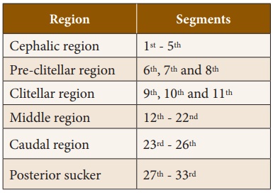
3. Divisions of the Body
The body of leech is
divided into six regions.
4. Body wall
Body wall of leech
includes five layers:
(i) cuticle (outermost
layer) (ii) epidermis which lies below the cuticle (iii) dermis
which lies below the epidermis formed of connective tissue (iv) muscular
layer formed of circular and longitudinal muscles (v) botryoidal tissue
lies beneath longitudinal muscles and fills the entire coelom around the gut.
5. Locomotion
Locomotion in leech
takes place by (i) looping or crawling movement (ii) Swimming movement.
(i) Looping or Crawling movement
This type of movement is
brought about by the contraction and relaxation of muscles. The two suckers
serve for attachment during movement on a substratum.
(ii) Swimming movement
Leeches swim very
actively and perform undulating movements in water.
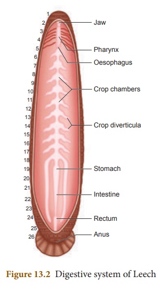
6. Digestive System
The digestive system
includes the long alimentary canal and the digestive glands.
Alimentary Canal
The alimentary canal of
leech is a straight tube running from the mouth to the anus. Mouth is a
triradiate aperture situated in the middle of the anterior sucker
that leads into the small buccal cavity. The wall of the buccal cavity bears
three jaws with single row of minute teeth. The jaws are provided with papillae
which bear the openings of salivary glands. Mouth and buccal cavity
occupy the first five segments.
The buccal cavity leads
into muscular pharynx. It is surrounded by salivary glands. The
secretion of saliva contains hirudin which prevents the coagulation of
blood. Pharynx leads into crop through a short and narrow oesophagus.
Crop is the largest portion
of the alimentary canal. It is divided into a series of 10 chambers. The
chambers communicate with one another through circular apertures surrounded by sphincters.
A pair of lateral, backwardly directed caecae arises as blind
outgrowth from each chamber known as caeca or diverticula. Crop
and its diverticula can store large amount of blood which can be slowly
digested.
The last chamber of crop
opens into stomach. The stomach leads into intestine which is a small
straight tube that opens into rectum. The rectum opens to the exterior
by anus.
Food, Feeding and Digestion
The leech feeds by
sucking the blood of cattle and other domestic animals. During feeding the
leech attaches itself to its victim strongly by the posterior sucker. The leech
makes a triradiate or Y shaped incision in the skin of the host
by the jaws protruded through the mouth. The blood is sucked by muscular
pharynx and the salivary secretion is poured.
The ingested blood is
stored in crop chambers and its diverticulum. The blood passes from the crop
into the stomach. Digestion takes place in stomach by the action of proteolytic
enzyme. The digested blood is then absorbed slowly by the intestine. Undigested
food is stored in rectum and egested through anus.
Leeches prevent blood
clotting by secreting a protein called hirudin. They also inject an
anaesthetic substance that prevents the host from feeling their bite.
7. Respiratory System
Respiration takes place
through the skin in leech. Dense network of tiny blood vessels called as
capillaries containing the haemocoelic fluid extend in between the cells
of the epidermis. The exchange of respiratory gases takes place by diffusion.
Oxygen dissolved in water diffuses through the skin into haemocoelic fluid,
while carbon dioxide diffuses out. The skin is kept moist and slimy due to
secretion of mucus which also prevents it from drying.
8. Circulatory System
In leech, circulation is
brought about by haemocoelic system. There are no true blood vessels.
The blood vessels are replaced by channels called haemocoelic channels
or canals filled with blood like fluid. The coelomic fluid
contains haemoglobin.
There are four
longitudinal channels. One channel lies above (dorsal) the alimentary canal,
one below (ventral) the alimentary canal. The other two channels lie on either
(lateral) side of the alimentary canal which serve as heart and have inner
valves. All the four channels are connected together posteriorly in the 26th
segment.
9. Nervous System
The central nervous system of leech consists of a nerve ring and a paired ventral nerve cord.
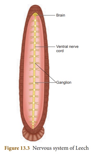
The nerve ring surrounds the pharynx and is formed of suprapharyngeal
ganglion (brain), circumpharyngeal connective and subpharyngeal
ganglion. The subpharyngeal ganglion lies below the pharynx and is
formed by the fusion of four pairs of ganglia.
10. Excretory System
In leech, excretion
takes place by segmentally arranged paired tubules called nephridia.
There are 17 pairs of nephridia which open out by nephridiopores
from 6th to 22nd segments.
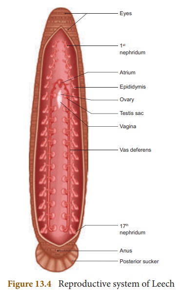
11. Reproductive System
Leech is hermaphrodite
because both the male and female reproductive organs are present in the same
animal.
Male Reproductive System
There are eleven pairs
of testes, one pair in each segment from 12 to 22 segments. They are in the
form of spherical sacs called testes sacs. From each testis
arises a short duct called vas efferens, which join with the vas
deferens. The vas deferens becomes convoluted to form the epididymis
or sperm vesicle, to store spermatozoa.
The epididymis leads to
a short duct called ejaculatory duct. The ejaculatory ducts on both
sides join to form the genital atrium. The atrium consists of two
regions, the coiled prostate glands and the penial sac consisting of penis that
opens through the male genital pore.
Female Reproductive System
It consists of ovaries,
oviducts and vagina. There is a single pair of ovary in the 11th segment on the
ventral side. Each ovary is a coiled ribbon-shaped structure.
The ova are budded off
from the ovary. From each ovary runs a short oviduct. The oviducts
of the two sides joins together, to form a common oviduct. The common oviduct
opens into a pear-shaped vagina which lies mid-ventrally in the
posterior part of the 11th segment.
Development
(i) Internal fertilization takes place. This is followed by cocoon
formation. Cocoon is also known as egg case which is formed
around the 9th, 10th and 11th segments.
(ii) Development is direct and proceeds in cocoon which contain
one to 24 embryos.
(iii) Young leech
resembling the adult emerges.
12. Parasitic Adaptations of Leech
Leeches lead a parasitic
mode of life by sucking the blood of vertebrates and show several important
adaptations in their structure.
a)
Blood is sucked by pharynx.
b)
Anterior and posterior ends of the body are provided with suckers
by which the animal attaches itself to the body of the host.
c)
The three jaws inside the mouth, causes a painless Y-shaped wound
in the skin of the host.
d)
The salivary glands produce hirudin which does not allow the blood
to coagulate. Thus, a continuous supply of the blood is maintained.
e)
Parapodia and setae are completely absent
f)
Blood is stored in the crop. It gives nourishment to the leech for
several months. Due to this reason there is no elaborate secretion of the
digestive juices and enzymes.
Related Topics