Chapter: The Massage Connection ANATOMY AND PHYSIOLOGY : Reproductive System
The External Genitalia - Female Reproductive System
THE EXTERNAL GENITALIA
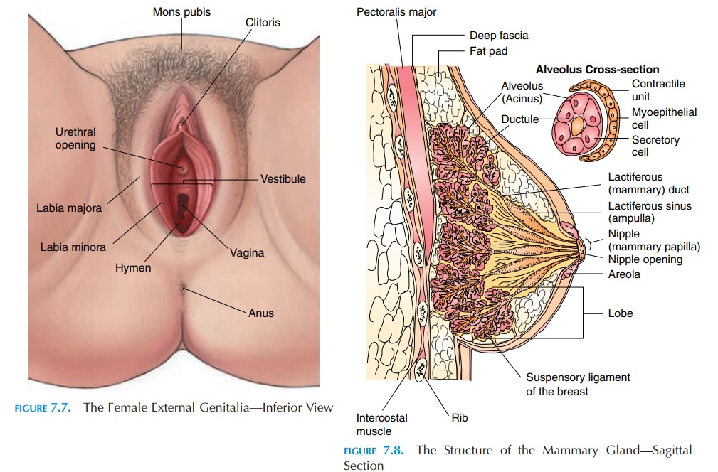
The region of the external genitalia (see Figure 7.7) is known as the vulva or pudendum. Adipose tissue over the pubic symphysis produces a bulge known as
Folds of skin cover the sides of the openings; the outermost thicker fold, with coarse hair (in adults), is known as the labia majora.Inner to this is the thinner, smooth, hairless fold known as the labia minora. The space enclosed by the labia minora is known as thevestibule. There are three major openings in the perineum. The most anterior opening is the urethra, with the vaginal opening pos-terior to it. The anal opening is the most posterior opening. Superior to the urethral opening is the cli-toris, the structure that is embryologically equivalentto the male penis. The clitoris is a small cylindrical mass of tissue that is erectile. As in males, it is capa-ble of enlarging in size when stimulated.
Glands in the External Genitalia Region
Many glands are found in this region that help keep the vestibule moist and provide lubrication. The pa-raurethral or Skene’s glandsare located near theurethral opening and the lesser and greater vestibu-lar (Bartholin’s) glands are found near the vaginal entrance. In addition, sebaceous and sweat glands lo-cated in the labia majora help with the function.
Breasts
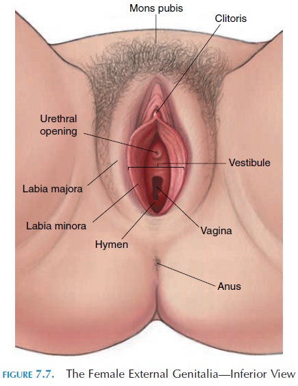
The glandular portion of the breast is known as the mammary gland. The two mammary glands are mod-ified sudoriferous (sweat) glands. The breasts are posi-tioned over the second to sixth ribs and lie superficial to the pectoralis major, part of the serratus anterior and anterior oblique muscles. Medially, the breasts ex-tend to the lateral margin of the sternum. Laterally, the breasts follow the anterior border of the axilla. The ax-illary process of the breast extends upwards and lat-erally toward the axilla close to the axillary vessels.
In nonpregnant individuals, the breasts (see Figure 7.8) are largely made up of fat and connective tissue, scattered with immature glands. During pregnancy, under the influence of many hormones, the glands en-large and occupy the major portion of the breast. The glands consist of 15 to 20 compartments, or lobes, which are separated by fat tissue. Between the lob-ules, suspensory ligaments(Cooper ligaments) ex-tend from the skin to the deep fascia over the pectoralis major, supporting the breast. Each lobe has many smaller compartments (lobules) that are col-lections of milk-secreting glands known as alveoli. The alveoli are lined by secretory cells and sur-rounded by spindle-shaped smooth muscle cells called myoepithelial cells. Contraction of these cells helps expel the milk toward the nipples. The alveoli open into larger ducts, which join with more ducts and eventually form about 15 largelactiferous ducts that open out through the nipple. Just before the ducts open into the nipple, they are enlarged into the lactiferous sinus (ampulla) where a larger volume ofmilk can be held before they are expelled. The nipple is a cylindrical part containing some erectile tissue. Surrounding the nipple is a circular pigmented area known as the areola. The color of the areola varies ac-cording to the complexion of the woman. During pregnancy, the areola becomes darker and enlarged in area.
Related Topics