Chapter: The Massage Connection ANATOMY AND PHYSIOLOGY : Skeletal System and Joints
The Elbow Joint
THE ELBOW JOINT
Articulating Surfaces and Type of Joint
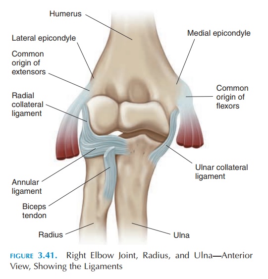
The elbow joint (see Figure 3.41) is a hinge joint with three components. The humeroulnar joint is where the trochlea of the humerus articulates with the trochlear notch of the ulna. The humeroradial joint is formed by the capitulum of the humerus and the head of the radius, and the proximal radioulnarjoint is the articulation between the head of the ra-dius and the radial notch of the ulna. The latter is not part of the hinge but is a pivot joint. The capsule and joint cavity are continuous for all three joints. The el-bow joint is relatively stable because it is well sup-ported by bone and ligaments.
Ligaments
Two major ligaments—the ulnar (medial) collateral ligament and the radial (lateral) collateral liga-ment— support the joint on either side and rise from the medial and lateral epicondyle of the humerus, re-spectively. The head of the radius is held in the radial notch of the ulna by the annular ligament.
Bursa
An olecranon bursa is located posteriorly over the olecranon process.
Possible Movements
The elbow joint allows flexion and extension. Fore-arm supination and pronation are also possible and a result of the articulation between the radius and ulna proximally and distally.
Range of Motion
Flexion, 135°
Extension, 0–5°
Supination, 90°
Pronation, 90°
Muscles
Muscles that flex the elbow:
Primary flexors
Brachialis Biceps brachii
Secondary flexors
Brachioradialis
Supinator
Muscles that help with extension:
Primary extensor
Triceps
Secondary extensor
Anconeus
Muscles that help with supination:
Primary supinators
Biceps
Supinator
Secondary supinator
Brachioradialis
Muscles that help with pronation:
Primary pronators
Pronator teres
Pronator quadratus
Secondary pronator
Flexor carpi radialis
Physical Assessment
Inspection
Note the angle made by the forearm with the upper arm—the carrying angle. Normally, it is about 5° in men and 10–15° in women. Swelling, scars, and skin discolorations should be recorded.
Palpation
The bony prominences that can be easily felt at the el-bow are the medial epicondyle, the olecranon, the olecranon fossa of the humerus into which the ole-cranon fits, the ulnar border, the lateral epicondyle, and the head of the radius.
Medially, the ulnar nerve can be easily located in the sulcus between the medial epicondyle and the olecranon process. If the olecranon bursa is inflamed, it can be felt as a thick and boggy structure over the olecranon. Tenderness over the lateral collateral liga-ment and the annular ligament can be identified.
The shallow depression in front of the forearm is the cubital fossa. The biceps tendon and the pulsa-tion of the brachial artery can be felt here.
In addition, the various muscles, active and pas-sive range of motion should be checked.
Related Topics