Chapter: The Massage Connection ANATOMY AND PHYSIOLOGY : Skeletal System and Joints
The Ankle Joint and Joints of the Foot
THE ANKLE JOINT AND JOINTS OF THE FOOT
Articulating Surfaces and Type of Joint
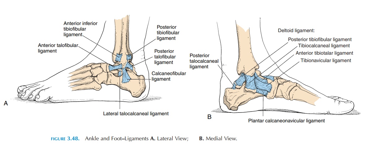
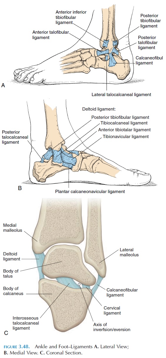
The ankle joint (see Figure 3.48) is formed by the dis-tal end of the tibia, fibula, and the superior surface of the talus. This joint is also known as the talocrural joint. It is a hinge joint with the lateral and medialaspect of the capsule thickened to form ligaments.
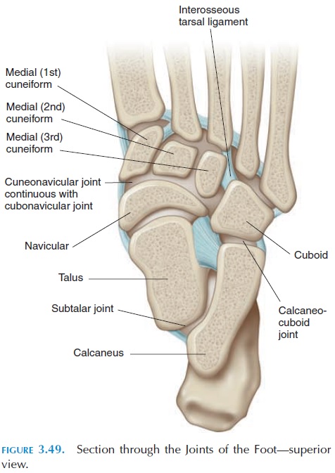
Other articulations (see Figure 3.49) occur between the talus and calcaneus (subtalar joint); between the tarsal bones (midtarsal joints) and talocalcaneonav-icular and calcaneocuboid joints; between the ante-rior tarsals (anterior tarsal joints) and the cubonav-icular , cuneonavicular, cuneocuboid, and intercuboid joints; between the tarsals and the metatarsals (tar-sometatarsal joints); between the metatarsal andphalanges (metatarsophalangeal joint); and be-tween the phalanges (the proximal anddistal inter-phalangeal joints).
Ligaments
The medial ligament, or the deltoid ligament, is a thickening of the medial fibrous capsule that attaches the medial malleolus to the navicular, calcaneus, and talus bones. The calcaneofibular ligament extends from the lateral malleolus to the calcaneus. Anteri-orly and posteriorly, ligaments extend from the lat-eral malleolus to the talus to form the anteriortalofibular (most frequently injured) and posterior talofibular ligaments. The various ligaments pre-vent tilt and rotation of the talus and forward and backward movement of the leg over the talus.
Possible Movements
The ankle allows dorsiflexion and plantar flexion. However, the subtalar joint and tarsal joints allow further movement. Eversion and inversion is possible at the subtalar joint. The foot can be adducted and abducted at the midtarsal joints. The metatarsopha-langeal joints and interphalangeal joints are hinge joints, allowing flexion and extension of the toes.
Range of Motion
Dorsiflexion, 20°
Plantar flexion, 50°
Inversion and eversion, 5°
Adduction, 20°
Abduction, 10°
Flexion (toes), 45°
Extension, 70–90°
Muscles
Muscles that cause plantar flexion:
Primary plantar flexors
Gastrocnemius
Soleus
Secondary plantar flexors
Tibialis posterior
Flexors of the toes
Peroneus longus and brevis
Muscles that cause dorsiflexion:
Tibialis anterior
Peroneus tertius
Extensors of the toes
Muscle that inverts the foot:
Tibialis anterior and posterior
Muscle that everts the foot:
Peroneus longus, brevis, and tertius
Physical Assessment
Inspection
The external appearance of the shoe and foot should provide information. The alignment of the toes and the shape of the foot and arches should be inspected. The color of the skin and presence of swelling should also be noted.
Palpation
The bones of the foot and ankle are easily palpated. Some bony prominences that can be located are the malleoli, talus, calcaneus, and the metatarsal and phalanges. The deltoid ligament is also palpable infe-rior to the medial malleolus. The long saphenous vein, if dilated may be visible just anterior to the me-dial malleolus. Both active and passive range of mo-tion should be tested at the various joints.
Related Topics