Chapter: Microbiology and Immunology: Mycology, Fungi: Superficial, Cutaneous, and Subcutaneous Mycoses
Superficial Mycoses
Superficial Mycoses
Superficial mycosis caused by different fungi is restricted to the outermost layers of the skin and hair. The condition usually causes cosmetic problem, which can be easily diag-nosed and treated. It includes four important conditions: (a) pityriasis versicolor, (b) tinea nigra, (c) black piedra, and (d) white piedra.
Pityriasis Versicolor
Pityriasis versicolor or tinea versicolor is a superficial infection of the skin caused by Malassezia furfur (Pityrosporum orbiculare). M. furfur requires fatty acids for growth, hence is cultured onthe Sabouraud’s dextrose agar (SDA) overlayed with a layer of olive oil. On incubation at 37°C, the fungus produces creamy colonies within 5–7 days. The lactophenol cotton blue (LPCB) wet mount of these colonies shows budding yeast cell along with a number of bottle-shaped cells. The fungus is found in parts of the body rich in sebaceous glands. The lesions of pity-riasis versicolor are found most commonly on the upper tissue, arms, and abdomen. They appear as hypopigmented macu-lar lesions often associated with slight scaling or itching. The condition is mostly asymptomatic. It occurs most frequently in hot and humid weather.
Laboratory diagnosis of the condition is usually made by demonstration of both budding yeast cell and hyphae in KOH preparation of skin scrapings. Characteristic “spaghetti and meatballs” appearance of fungus is demonstrated in the microscopy of KOH preparation of the skin. Culture is not carried out routinely for diagnosis. Topical miconazole is treatment of choice.
Tinea Nigra
Tinea nigra is an infection of keratinized layer of skin caused by Exophiala werneckii or Cladosporium werneckii. C. werneckii is a dimorphic fungus that produces melanin. The fungus on the SDA grows as yeast with many cells in various stages of cell divi-sion producing typical two-celled oval structure, on primary isolation from clinical specimen. On prolonged incubation, elongated hyphae develop and in older cultures, mycelia and conidia are predominantly found.
A well-demarcated brown-black macular lesion, which appears as brownish spot of the skin, is typical manifestation of the condition. These brownish to black lesions are most commonly seen on palms and soles.
Laboratory diagnosis of tinea nigra is made by microscopy of the KOH preparation of skin scrapings collected from the affected part. Typical darkly pigmented yeast-like cells and hyperfragmented hyphae are demonstrated. Culture of the skin scraping on the SDA confirms the diagnosis.
Black Piedra
Black piedra is a superficial infection of the hair caused by Piedraiahortae, a dematiaceous fungus. The fungus occurs in the perfectstate when it colonizes the shaft of hairs. Culture of specimens on SDA shows slow-growing brown to reddish black mycelium, which is considered asexual or anamorphic stage of the fungus. The teleomorphic state, which is the perfect state of the fungus, is occasionally found in old cultures. At this state asci, which contain spindle-shaped ascospores, develop within specialized structures.
Infection of shaft of hairs of beard and scalp is the major clinical feature of black piedra. Laboratory diagnosis of the condition is made by demonstration of nodules containing asci with spindle-shaped ascospores in 10% KOH mount of the hair.
White Piedra
White piedra is an infection of the hair caused by yeast-like organism Trichosporon beigelli. The fungus can be grown on SDA and other media containing cycloheximide. On SDA, it forms green-colored colonies, which subsequently become yellow-ish gray and wrinkled. Microscopic examination of the colony shows septate hyphae that break rapidly to form arthroco-nidia. The latter subsequently become round and develop to blastoconidia.
White piedra is commonly found in South America, Central and Eastern Europe, and Japan.
The hair of scalp, moustache, and beard are commonly affected in white piedra. The development of a soft, pasty, cream-colored growth along infected hair shaft characterizes the condition. The initial growth of T. beigelli occurs beneath the epidermis of hair. The infected hair shaft consists of mycelium that rapidly fragments to arthroconidia.
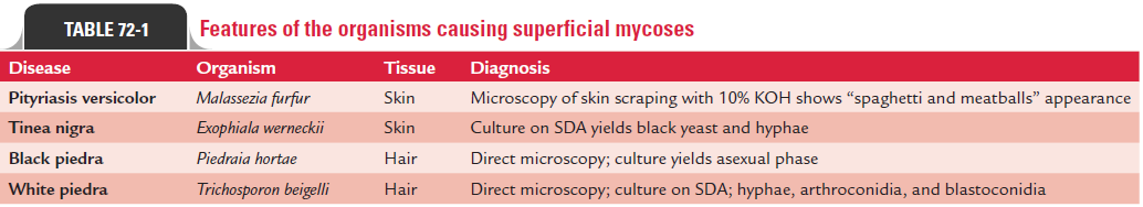
Laboratory diagnosis of the condition is made by demon-stration of fragmented hyphae that develop into arthroconidia or produce blastoconidia in 10% KOH mount of hair. Culture of the fungus from clinical specimen confirms the diagnosis. Features of the organisms causing superficial mycoses are summarized in Table 72-1.
Related Topics