Chapter: Human Neuroanatomy(Fundamental and Clinical): Sensory Receptors and Neuromuscular Junctions
Structure of a Typical Motor End Plate
STRUCTURE OF A TYPICAL MOTOR END PLATE:
In the region of the motor end plate axon terminals are lodged in grooves in the sarcolemma covering the sole plate. Between the axolemma (over the axon) and the sarcolemma (over the muscle fibre) there is a narrow gap (about 40 nm) occupied by various proteins that form a basal lamina. It follows that there is no continuity between axoplasm and sarcoplasm.
Axon terminals are lodged in grooves in the sarcolemma covering the sole plate. In section (Fig. 4.5) this groove is seen as a semicircular depression. This depression is the primary cleft. The sarcolemma in the floor of the primary cleft is thrown into numerous small folds resulting in the formation of secondary (or subneural) clefts.
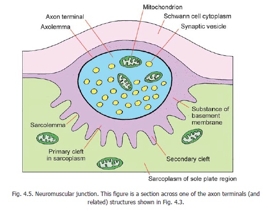
In the region of the sole plate the sarcoplasm of the muscle fibre is granular. It contains a number of nuclei and is rich in mitochondria, endoplasmic reticulum and Golgi complexes.
Axon terminals are also rich in mitochondria. Each terminal contains vesicles similar to those seen in presynaptic boutons. The vesicles contain the neurotransmitter acetyl choline. Acetyl choline is released when nerve impulses reach the neuromuscular junction. It initiates a wave of depolarisation in the sarcolemma resulting in contraction of the entire muscle fibre. Thereafter the acetyl choline is quickly destroyed by the enzyme acetyl choline esterase. The presence of acetyl choline receptors has been demonstrated in the sarcolemma of the sole plate.
Nerve endings on smooth muscle
Nerve fibres innervating smooth muscle are unmyelinated. They end a short distance away from the myocyte surface. (In other words axolemma and sarcolemma do not come into contact). At most places, the nerve fibres are covered by Schwann cell cytoplasm. However, at places this cytoplasm is retracted exposing a segment of the axon. This segment of the axon shows the presence of vesicles. Neurotransmitter released from the vesicles diffuses to the myocytes.
In sympathetic terminals the vesicles contain catecholamines (usually noradrenaline). Monamine oxidases present in relation to sympathetic endings destroy catecholamines and thus regulate sympathetic activity. At parasympathetic terminals the vesicles in axon terminals are clear. They contain acetyl choline.
Recently it has been shown that some autonomic terminals contain neither noradrenaline nor acetyl choline. These are described as non-adrenergic non-cholinergic endings. The neurotransmitter present at these endings is probably a purine (adenosine triphosphate). Such fibres have been demonstrated in the walls of the alimentary and urinary tracts, and also in the CNS. These endings are believed to be predominantly inhibitory.
Other effector endings:
Apart from muscle, effector endings are present in relation to glands (secretomotor endings), to myoepithelial cells, and to adipose tissue.
Related Topics