Chapter: Microbiology
Stains and staining reactions

STAINS AND STAINING REACTIONS
Bacteria are semi-transparent and consist of a clear proto-plasmic matter that differs slightly in refractive index from the medium in which they are growing. It is difficult to observe the bacteria in unstained state, except when special methods of illu-mination are used, to see them in the unstained state.
Stains are useful for the following reasons.
l It makes the microscopic semi-transparent objects visible
l To study the shape and size
l To reveal the presence of various internal and external struc-tures
l To produce specific chemical and physical reaction
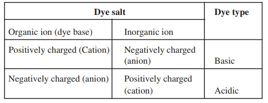
The term stain and dye are not the same. A colouring agent that is used for general purposes is called a dye. The one that is used for biological purposes is called a stain. Based on their chemi-cal behavior, the dyes are classified as acidic, basic and neutral.
An acid (or anionic) dye has a negative charge. eg., Eosin, Rose Bengal and Acid fuchsin. The negatively charged groups are carboxyls (-COOH) and Phenolic hydroxyls (-OH). Since they are negatively charged, bind to positively charged cell structures. pH plays an important role in the effectiveness of staining, because the nature and the degree of the charge on cell components change with pH. The anionic dyes stain better under acidic conditions, where the proteins and many other molecules carry a positive charge.
A basic dye (or cationic) carries a positive charge. eg., Methylene Blue, basic fuchsin, crystal violet, malachite green, safranin. Ba-sic dyes bind to negatively charged molecules like nucleic acid and many proteins. Since the bacterial cells surfaces are negatively charged, basic dyes are most often used in Bacteriology. Basic dyes are normally available as chloride salts.
A neutral dye is a complex salt of a dye acid with a dye base.
The dyes used in bacteriology have two features in common.
l They have chromophore groups, groups with double bonds, that give the dye its colour
l They can bind with cells by ionic, covalent or hydrophobic bonding.
Relationship between the type of the dye and its charge when dissociated is summarized.
In positive staining procedure, a stain that has a positively gedchromophorear (coloured portion of the stain molecule) is attracted to the negatively charged outer surface of the microbial cell. A stain such as Methylene blue has a positively charged blue portion of the molecule that stains the microorganism.
In negative staining procedures, a negatively charged chro-mophore is repelled by the negatively charged microorganisms, resulting in negative or indirect staining of the microbial cell. Ni-grosin and Indian ink are frequently used for negative staining of microbial cells, and this type of staining is particularly useful for viewing some structures such as capsules that surround some bac-terial cells.
Stains are generally prepared largely as aqueous solutions. However in some cases stock solutions are prepared in alcohol, and are diluted with water as needed. Since alcohol removes the stains, pure alcoholic solutions should not be used. Staining solu-tions are prepared to contain low concentrations of stains rarely exceeding 1%. A very dilute staining solution activity for a long period of time will produce much better results then a more con-centrated solution acting for a shorter interval. This procedure has to be followed to reveal internal structure in bacteria.
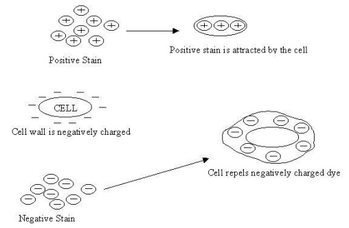
Figure:3.1 The interaction of a cell with negative and positive stain re-agents: The outer layer of a cell is negatively charged and a positive stain is attracted to the cell, whereas a negative stain is repelled.
Staining reactions-Interpretation notes
When the pH of the surroundings of the microbial cells is either neutral or alkaline, all microbial cells have a negative charge on their surface, called the surface charge. Many bacterial cultures produce acids, thereby adding hydrogen ions to a culture medium and decreasing it pH. These hydrogen ions(H+) interact with the surface of the negative charges on the surface. When this happens, the cell surface no longer strongly attracts positively charged dye ions (basic dyes). Thus the microbes from acidic environments stain poorly with basic dyes. For this reason, the basic dyes are made up as alkaline solutions. For example, potassium hydroxide (KOH) is added to solutions of methylene blue to form the called Loeffler’s Methylene blue.
Some bacteria excrete alkaline materials during growth and this decreases the number of available hydrogen ions in the cul-ture medium. Under such conditions, the cell surface has a greater negative charge, which is more attractive to basic dyes and there- fore, allows greater binding, penetration and internal staining of the microbe. Basic dyes stain microorganisms better under neutral or alkaline conditions.
If the dye base molecule has a negative charge, it is repelled by the cell’s negativelyThusnegativelycharged charsurface. ged dyes neither bind to the cell’s surface nor are they able to penetrate into the cell. These are called acid dyes.
The methodology for using acid dyes are different from ba-sic dyes. An acid dye is mixed with a drop of culture smeared on a microscope slide and allowed to air dry. The negatively charged cells are not stained by the negatively charged dye, and they ap-pear as clear area surrounded by a coloured back ground. Nega-tively charged dyes used in this way are called negative stains.
Under neutral or alkaline conditions, the negative stains (acidic dyes) work better, because these conditions allow the sur-face charge to be more negative. Negative stains are of limited usefulness for those using light microscopes, but they can be used to avoid some of the disadvantages of staining with basic dyes.
Simple staining
A simple staining solution, contains only one stain, which is dissolved in a solvent. It is applied to the microorganism in one application. The microorganisms give the colour characteristic of the staining solution. The purpose of simple staining is to reveal the size and shape of the microorganism. The simple stains that are commonly used by the microbiologists for routine purposes are dilute solution of carbol fuchsin, crystal violet and methylene blue.
Methylene blue is more frequently used than any other stain in bacteriology. It is because of its strong nature and it stains nu-clei and nucleic acid granules very intensively. Methylene blue is used for the rapid survey of bacterial population of milk. It is also used for the diagnosis of Diphtheria. This stain is incorporated with Eosin in Lactose agar to distinguish typicalinconE-.coli taminated water.
Differential Staining
In this procedure, more than one dye is employed. Differen-tial staining procedure helps to divide the bacteria into separate groups based on staining characteristics. The two most important differential stains used by bacteriologists are Gram stain and Acid-fast stain.
Gram Staining
The simple staining procedure makes to visualize bacteria clearly, but it does not distinguish between organisms of similar morphology. In 1884, a Danish Physician named, Christian Gram discovered a new technique to differentiate the bacteria of similar morphology. He used two dyes in sequence, each of a different colourganisms.Thethatorretain the colour of the first dye are called Gram positive and those that cannot retain the first dye when washed with a decolourizing solution, but then take on the colour of the second dye are called Gram negative . In this method, the fixed bacterial smear is subjected to the following staining regents in the order of sequence listed below:
Crystal violet - - > Iodine solution - - > alcohol (decolourizing agent) - - > Safranin.
Principle
The Gram-positive bacteria will retain the crystal violet and appear deep violet in colour. The Gram-negative bacteria lose the crystal violet on decolorization and are counter stained by the sa-franine and appear red in colour. Iodine solution is used as a mor-dant that fixes the primary stain in or on a substrate by combining with the dye to form an insoluble compound-mordant, for the first stain.
The exact mechanism of action of this staining technique is not clearly understood. However, the most plausible explanations for the reactions are associated with the structure and composition of the cell wall.
The cell walls of Gram-negative bacteria are thinner than that of Gram-positive bacteria and contain a higher percentage of lipid content. During the staining of Gram-negative bacteria, the alcohol treatment extracts the lipid. This results in increased po-rosity or permeability of the cell wall. The crystal violet-iodine (CV-I) complex, thus can be extracted and the Gram-negative bac-teria is decolorized. The cells subsequently take up the colour of the counter stain safranin.
The cell walls of Gram-positive bacteria with lower lipid content become dehydrated during alcohol treatment. The pore size decreased, permeability is reduced and the CV-I complex can-not be extracted. Therefore, the Gram-positive cells remain purple-violet.
Endospore Staining
Endospore formation is a distinguishing feature of the fam-ily Bacillaceae, which includes members of the aerobic genus, and the Bacillusanaerobic genus, Clostridium. Endospore resists adverse environmental conditions such as dryness, heat and poor nutrient supply. The endospore is a highly retractile body formed within the vegetative bacterial cell at a certain stage of growth.The size, shape, and position of the spore are relatively constant characteristics of a given species and are therefore, of some value in distinguishing the kind of bacillus from another. The position of the spore in the cell may be central, sub terminal or terminal. It may be the same diameter as the cell, smaller, or larger causing a swelling of the cell.
Endospores strongly resist application of simple dyes, but once stained are quiet resistant to decolorization. This character suggests one way to make the structure visible. If simple stains are used, the body of the bacillus is deeply colored, whereas the spore is unstained and appears as a clear area in the organism. By vigor-ous staining procedures the dye can be introduced into the sub-stance of the spore. When thus stained, the spore tends to retain the dye after treatment with decolorizing agents.
To make the distinction clear between the spore and the veg-etative portion of the cell, a contrasting counter stain is usually applied in the ordinary fashion and the resulting picture shows the initial stain taken up by the spore and the second stain appear in the cytoplasm. Thus, it makes for a very simple method of distin-guishing the endospore from the vegetative cell.
Related Topics