Chapter: Essentials of Anatomy and Physiology: The Nervous System
Spinal Cord
THE SPINAL CORD
The spinal cord transmits impulses to and from the brain and is the integrating center for the spinal cord reflexes. Although this statement of functions is very brief and sounds very simple, the spinal cord is of great importance to the nervous system and to the body as a whole.
Enclosed within the vertebral canal and the meninges, the spinal cord is well protected from mechanical injury. In length, the spinal cord extends from the foramen magnum of the occipital bone to the disc between the first and second lumbar vertebrae.
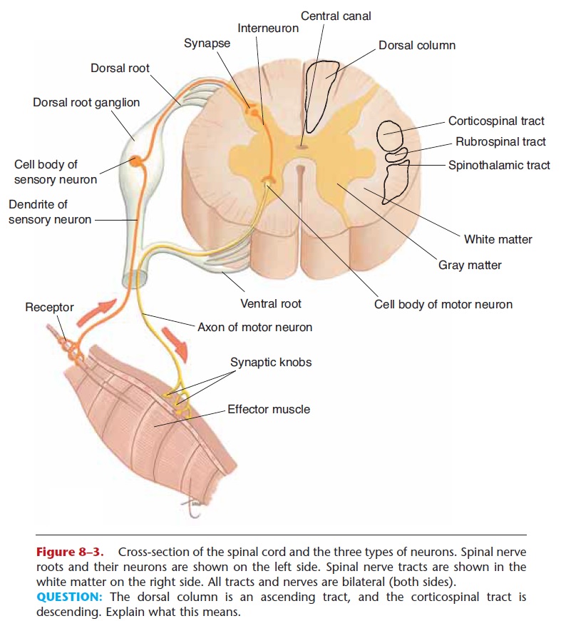
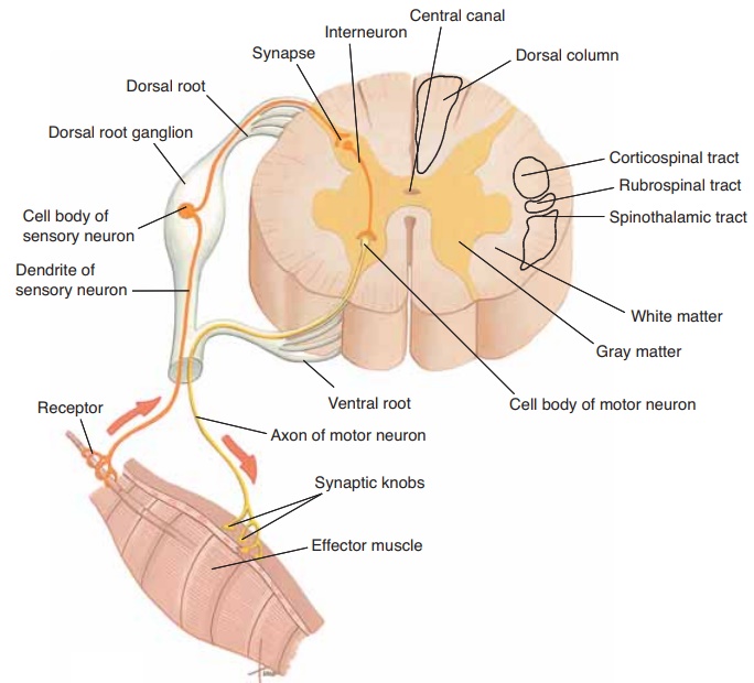
A cross-section of the spinal cord is shown in Fig. 8–3; refer to it as you read the following. The internal gray matter is shaped like the letter H; gray matter consists of the cell bodies of motor neurons and interneurons. The external white matter is made of myelinated axons and dendrites of interneurons. These nerve fibers are grouped into nerve tracts based on their functions. Ascending tracts (such as the dor-sal columns and spinothalamic tracts) carry sensory impulses to the brain. Descending tracts (such as the corticospinal and rubrospinal tracts) carry motor impulses away from the brain. Lastly, find the central canal; this containscerebrospinal fluid and is con-tinuous with cavities in the brain called ventricles.
SPINAL NERVES
There are 31 pairs of spinal nerves, those that emerge from the spinal cord. The nerves are named according to their respective vertebrae: 8 cervical pairs, 12 tho-racic pairs, 5 lumbar pairs, 5 sacral pairs, and 1 very small coccygeal pair. These are shown in Fig. 8–4; notice that each nerve is designated by a letter and a number. The 8th cervical nerve is C8, the 1st thoracic nerve is T1, and so on.
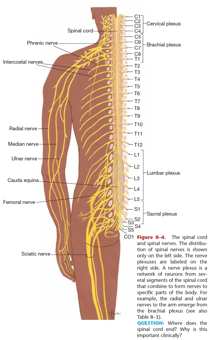
Figure 8–4. The spinal cord and spinal nerves. The distribu-tion of spinal nerves is shown only on the left side. The nerve plexuses are labeled on the right side. A nerve plexus is a network of neurons from sev-eral segments of the spinal cord that combine to form nerves to specific parts of the body. For example, the radial and ulnar nerves to the arm emerge from the brachial plexus (see also Table 8–3).
QUESTION: Where does the spinal cord end? Why is this important clinically?
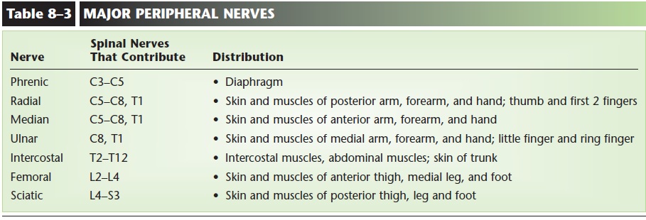
In general, the cervical nerves supply the back of the head, neck, shoulders, arms, and diaphragm (the phrenic nerves). The first thoracic nerve also con-tributes to nerves in the arms. The remaining thoracic nerves supply the trunk of the body. The lumbar and sacral nerves supply the hips, pelvic cavity, and legs. Notice that the lumbar and sacral nerves hang below the end of the spinal cord (in order to reach their proper openings to exit from the vertebral canal); this is called the cauda equina, literally, the “horse’s tail.” Some of the important peripheral nerves and their destinations are listed in Table 8–3.
Each spinal nerve has two roots, which are neurons entering or leaving the spinal cord (see Fig. 8–3). The dorsal root is made of sensory neurons that carry impulses into the spinal cord. The dorsal root gan-glion is an enlarged part of the dorsal root that con-tains the cell bodies of the sensory neurons. The term ganglion means a group of cell bodies outside the CNS. These cell bodies are within the vertebral canal and are thereby protected from injury.
The ventral root is the motor root; it is made of the axons of motor neurons carrying impulses from the spinal cord to muscles or glands. The cell bodies of these motor neurons, as mentioned previously, are in the gray matter of the spinal cord. When the two nerve roots merge, the spinal nerve thus formed is a mixed nerve.
SPINAL CORD REFLEXES
When you hear the term reflex, you may think of an action that “just happens,” and in part this is so. A reflex is an involuntary response to a stimulus, that is, an automatic action stimulated by a specific change of some kind. Spinal cord reflexes are those that do not depend directly on the brain, although the brain may inhibit or enhance them. We do not have to think about these reflexes, which is very important, as you will see.
Reflex Arc
A reflex arc is the pathway that nerve impulses travel when a reflex is elicited, and there are five essential parts:
1. Receptors—detect a change (the stimulus) and generate impulses.
2. Sensory neurons—transmit impulses from recep-tors to the CNS.
3. Central nervous system—contains one or more synapses (interneurons may be part of the pathway).
4. Motor neurons—transmit impulses from the CNS to the effector.
5. Effector—performs its characteristic action.
Let us now look at the reflex arc of a specific reflex, the patellar (or knee-jerk) reflex, with which you are probably familiar. In this reflex, a tap on the patellar tendon just below the kneecap causes extension of the lower leg. This is astretch reflex, which means that a muscle that is stretched will automatically contract. Refer now to Fig. 8–5 as you read the following:
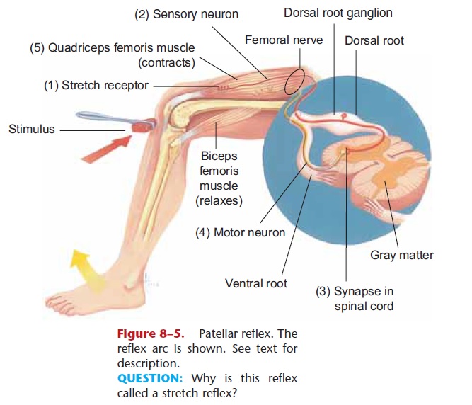
QUESTION: Why is this reflex called a stretch reflex?
In the quadriceps femoris muscle are (1) stretch receptors that detect the stretching produced by strik-ing the patellar tendon. These receptors generate impulses that are carried along (2) sensory neurons in the femoral nerve to (3) the spinal cord. In the spinal cord, the sensory neurons synapse with (4) motor neu-rons (this is a two-neuron reflex). The motor neurons in the femoral nerve carry impulses back to (5) the quadriceps femoris, the effector, which contracts and extends the lower leg.
The patellar reflex is one of many used clinically to determine whether the nervous system is functioning properly. If the patellar reflex were absent in a patient, the problem could be in the thigh muscle, the femoral nerve, or the spinal cord. Further testing would be needed to determine the precise break in the reflex arc. If the reflex is normal, however, that means that all parts of the reflex arc are intact. So the testing of reflexes may be a first step in the clinical assessment of neurologic damage.
You may be wondering why we have such reflexes, these stretch reflexes. What is their importance in our everyday lives? Imagine a person standing upright—is the body perfectly still? No, it isn’t, because gravity exerts a downward pull. However, if the body tilts to the left, the right sides of the leg and trunk are stretched, and these stretched muscles automatically contract and pull the body upright again. This is the purpose of stretch reflexes; they help keep us upright without our having to think about doing so. If the brain had to make a decision every time we swayed a bit, all our concentration would be needed just to remain standing. Since these are spinal cord reflexes, the brain is not directly involved. The brain may become aware that a reflex has taken place, but that involves another set of neurons carrying impulses to the brain.
Flexor reflexes (or withdrawal reflexes) are another type of spinal cord reflex. The stimulus is something painful and potentially harmful, and the response is to pull away from it. If you inadvertently touch a hot stove, you automatically pull your hand away. Flexor reflexes are three-neuron reflexes, because sensory neurons synapse with interneurons in the spinal cord, which in turn synapse with motor neurons. Again, however, the brain does not have to make a decision to protect the body; the flexor reflex does that automatically. The brain may know that the reflex has taken place, and may even learn from the experience, but that requires different neurons, not the reflex arc.
Related Topics